Polygalacturonases (PGs) catalyze the enzymatic depolymerization of pectates – polysaccharides that comprise the plant cell wall. Polymer disassembly of substrates by exo- and endo- PGs is carried out via a hydrolytic mechanism. Degradation of pectates in plant cell walls contributes to ripening of fruits, such as tomatoes and melons[1]. Microbial PGs have been identified to be a part of defense mechanisms because of their role in pathogen attack[2].
PG's are found in bacteria, fungi, plants, and animals. Plant PG's are involved in fruit ripening. Bacteria and fungal PG are involved in plant pathogenesis, often acting as plant virulence factors and involved in some of the initial pathogenic effects through their action of degrading the plant cell wall.
Function
Polygalacturonases hydrolyze α-(1-4) – glycosidic bonds between consecutive galacturonic acid residues in polygalacturonic acids. Structural variation has been identified among differing PGs depending on organismal origins and catalytic functions. For example, endo-polygalacturonases produced from Erwinia carotovora demonstrate functional similarity to pectate lyases in that they cleave polygalacturonic acids in a calcium-depended manner via β-elimination[2].
Not all PGs have ten coils in the parallel beta helix. They usually have approximately 10 coils.
Nomenclature for structural elements of the parallel beta helix
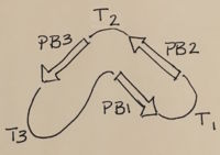
[3]. PB1 is Parallel Beta Sheet 1, T1 is Turn 1, between PB1 and PB2.,
PBH2 With the parallel beta helix fold, the three major beta sheets are call PB1, PB2, and PB3. The turns between strands are Turn 1 (T1) between PB1 and PB2, T2 is the turn between PB2 and PB3, and T3 is the turn between PB3 and PB1 of the next coil. For your reference, this is illustrated in the figure below.
3D Structures of polygalacturonase
Updated on 07-January-2018
References
- ↑ Hadfield KA, Bennett AB. Polygalacturonases: many genes in search of a function. Plant Physiol. 1998 Jun;117(2):337-43. PMID:9625687
- ↑ 2.0 2.1 Pickersgill R, Smith D, Worboys K, Jenkins J. Crystal structure of polygalacturonase from Erwinia carotovora ssp. carotovora. J Biol Chem. 1998 Sep 18;273(38):24660-4. PMID:9733763
- ↑ Yoder MD, Lietzke SE, Jurnak F. Unusual structural features in the parallel beta-helix in pectate lyases. Structure. 1993 Dec 15;1(4):241-51. PMID:8081738
[1]