This old version of Proteopedia is provided for student assignments while the new version is undergoing repairs. Content and edits done in this old version of Proteopedia after March 1, 2026 will eventually be lost when it is retired in about June of 2026.
Apply for new accounts at the new Proteopedia. Your logins will work in both the old and new versions.
Apoptotic protease-activating factor
From Proteopedia
| |||||||||||
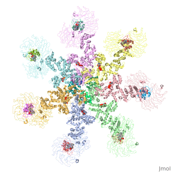
3D structures of apoptotic protease-activating factor-1
Updated on 15-March-2018
2ygs, 1cy5, 2p1h – hAPAF-1 CARD domain – human
1c15, 1cww – hAPAF-1 CARD domain – NMR
1z6t – hAPAF-1 + ADP
3ygs – hAPAF-1 CARD domain + procaspase 9
4rhw, 5wvc – hAPAF-1 CARD domain + caspase 9
3j2t, 3jbt – hAPAF-1 + cytochrome c
5juy, 5wve – hAPAF-1 + caspase 9 + cytochrome c
3sfz – mAPAF-1 + ADP - mouse
3shf – mAPAF-1 (mutant) + ADP
References
- ↑ Riedl SJ, Li W, Chao Y, Schwarzenbacher R, Shi Y. Structure of the apoptotic protease-activating factor 1 bound to ADP. Nature. 2005 Apr 14;434(7035):926-33. PMID:15829969 doi:10.1038/nature03465
- ↑ Furukawa Y, Sutheesophon K, Wada T, Nishimura M, Saito Y, Ishii H, Furukawa Y. Methylation silencing of the Apaf-1 gene in acute leukemia. Mol Cancer Res. 2005 Jun;3(6):325-34. PMID:15972851 doi:http://dx.doi.org/10.1158/1541-7786.MCR-04-0105
- ↑ Shalaeva DN, Dibrova DV, Galperin MY, Mulkidjanian AY. Modeling of interaction between cytochrome c and the WD domains of Apaf-1: bifurcated salt bridges underlying apoptosome assembly. Biol Direct. 2015 May 27;10:29. doi: 10.1186/s13062-015-0059-4. PMID:26014357 doi:http://dx.doi.org/10.1186/s13062-015-0059-4
