Vpr protein
From Proteopedia
| |||||||||||
3D Structures of Vpr protein
Updated on 03-April-2018
1m8l – Vpr - NMR - HIV-1
1esx - synthetic Vpr - NMR - HIV-1
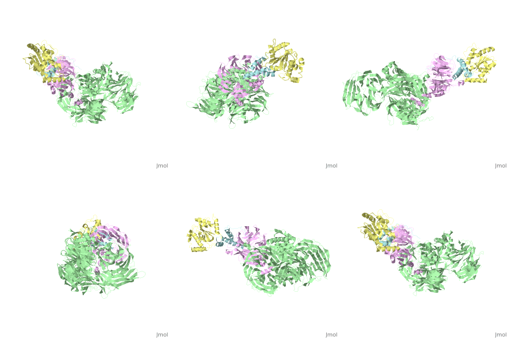
5jk7 – Vpr + DDB1 + DCAF-1 + UNG2 – X-ray solution - HIV-1
1x9v – Dimeric structure of the Vpr C-terminal domain - NMR
1vpc - C-terminal domain of Vpr - NMR - HIV-1
1fi0 - Vpr residues 13-33 in micelles - NMR - HIV-1
1bde - NMR solution of Vpr peptides connected to cell cycle arrest and nuclear provirus transfer
5b56 - Importin subunit alpha-1 + Vpr C-terminal domain - crystallographic analysis
1kzs, 1kzt, 1kzv - Vpr residues 34-51 - NMR - HIV-1
1dsj - Vpr residues 50-75 - NMR - HIV-1
1ceu - Vpr N-terminal domain - NMR - HIV-1
1dsk - Vpr residues 59-86 - NMR - HIV-1
4u1s - HLA-I + Beta-2-microglobulin + Vpr protein - X-ray diffraction

