Rhodopsin
Rhodopsin is a member of the G-protein coupled receptor (GPCR) family. Rhodopsin is the
common GPCR structure used to understand functionality of G-protein coupled receptors.
Rhodopsin is commonly found in the photoreceptors in the retina, specifically in the rod
photoreceptors and become activated by photons of light. Rhodopsin contains a chromophore
(compound that absorbs light), specifically 11-cis-retinal, which when active recruites G proteins
to transmit a signaling cascade in neural impulses to the gray matter of the occipital lobe. Once
the receptor has been activated, a new rhodopsin needs to be regenerated. Rhodopsin is
located in the rod outer segment (ROS) which consists of stacked disks enclosed by a
membrane. The entire family of GPCR’s have the common structure of seven alpha-helices
across membranes. Rhodopsin’s structure changes upon photoactivation. The sixth helix bends
away from the seventh creating a pocket that allows for binding of a G protein. A salt bridge
covers this pocket until the helices shift away from one another. Once the G protein has bound
then the signal can be transmitted to the occipital lobe. Over 120 point mutations to rhodopsin
have been identified which can lead to night blindness and more several visual problems[1].
Function
Rhodopsin performs two functions. One function is to bind retinal. Another function is to function as a G protein-coupled receptor. Rhodopsin is a protein that is essential for vision, especially in dim light. The photoreceptors in the retina that contain rhodopsin are rods. Rhodopsin is attached to 11-cis retinal which becomes excited by a photon of light. This excitation activates rhodopsin and leads to depolarizing of neurons. The depolarizing of neurons is how the image is transmitted to the brain. (https://ghr.nlm.nih.gov/gene/RHO#)
Disease
Mutations to rhodopsin can lead to stationary night blindness or retinitis pigmentosa.
Autosomal Dominant Congenial Stationary Night Blindness
Stationary night blindness is an autosomal dominant disease that causes a loss of vision in dim light. The mutation to rhodopsin causes the protein to be constantly activated. Since the protein is continuously activated without any photon of light, the brain continues to receive stimulation from the photoreceptors. The brain begins to ignore the visual stimulation from the rods, therefore leading to night blindness.
Retinitis Pigmentosa
Retinitis pigmentosa is also an autosomal dominant disorder, but can also be recessive in rare circumstances. A mutation that affect rhodopsin that cause retinitis pigmentosa result in a misfolding or transportation of the protein. Another mutation to rhodopsin can affect the activation of the protein in response to light. These mutations can lead to apoptosis of rods in the retina. Without rods to perceive dim light, night blindness results.
(https://ghr.nlm.nih.gov/gene/RHO#)
Relevance
Rhodopsin was the only GPCR that had a high-resolution crystal structure and was the basis for other GPCR structures. (https://www.ncbi.nlm.nih.gov/pubmed/24041646)
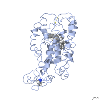
Structural highlights
This is rhodopsin without 11-cis retinal bound.
This is 11-cis retinal that rhodopsin binds.
This is rhodopsin with 11-cis retinal bound.
</StructureSection>
References
- ↑ Zhou XE, Melcher K, Xu HE. Structure and activation of rhodopsin. Acta Pharmacol Sin. 2012 Mar;33(3):291-9. doi: 10.1038/aps.2011.171. Epub 2012, Jan 23. PMID:22266727 doi:http://dx.doi.org/10.1038/aps.2011.171