Main Page

From Proteopedia

Revision as of 12:31, 18 October 2018 by Jaime Prilusky (Talk | contribs)
Jump to: navigation, search

Because life has more than 2D, Proteopedia helps to understand relationships between structure and function. Proteopedia is a free, collaborative 3D-encyclopedia of proteins & other molecules. ISSN 2310-6301

Selected Pages Art on Science Journals Education

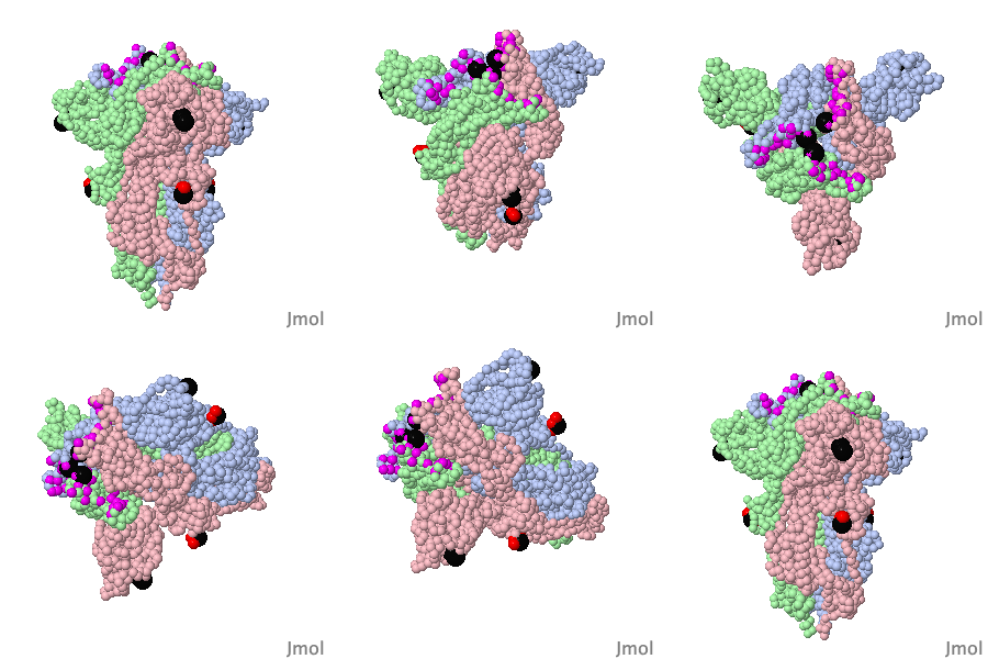
Lifecycle of SARS-CoV-2

What happens if a SARS-CoV-2 coronavirus enters your lung? This molecular animation visualises how the virus particle can take over the host cell and turns it into a virus factory. Eventually, the host cell produces so many viral particles that it dies and releases numerous new virus particles. >>> Visit this page >>>

About this image
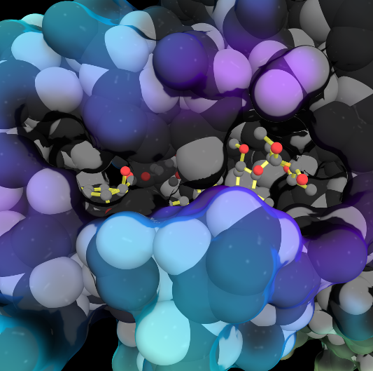
Opening a Gate to Human Health

by Alice Clark (PDBe)
In the 1970s, an exciting discovery of a family of medicines was made by the Japanese scientist Satoshi Ōmura. One of these molecules, ivermectin, is shown in this artwork bound in the ligand binding pocket of the Farnesoid X receptor, a protein which helps regulate cholesterol in humans. This structure showed that ivermectin induced transcriptional activity of FXR and could be used to regulate metabolism.

>>> Visit this page >>>

About this image
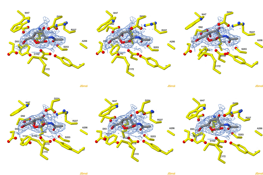
Geobacter nanowire structure surprise.

F Wang, Y Gu, JP O'Brien, SM Yi, SE Yalcin, V Srikanth, C Shen, D Vu, NL Ing, AI Hochbaum, EH Egelman, NS Malvankar. Cell 2019 doi: 10.1016/j.cell.2019.03.029
Bacteria living in anaerobic environments (no oxygen) need alternative electron acceptors in order to get energy from their food. An acceptor abundant in the earth's crust is red iron oxide ("rust"), which gets reduced to black iron oxide (magnetite). Many bacteria, such as Geobacter, get their metabolic energy by transferring electrons to acceptors that are multiple cell diameters distant, using protein nanowires. These were long thought to be pili. But when the structure of the nanowires was solved in 2019, to everyone's surprise, they turned out to be unprecedented linear polymers of multi-heme cytochromes. The hemes form an electrically conductive chain in the cores of these nanowires.

>>> Visit I3DC Interactive Visualizations >>>

About this image
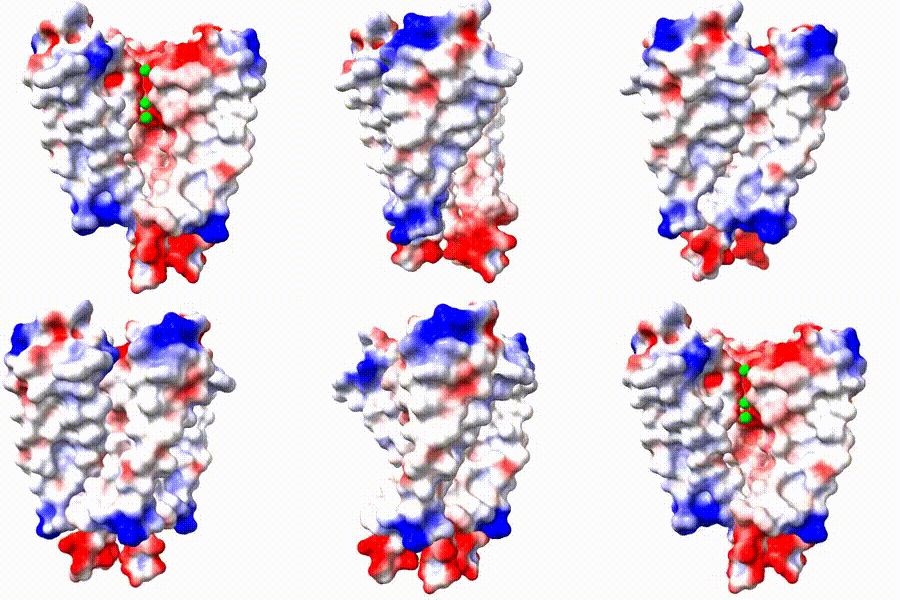
Make Your Own Electrostatic Potential Maps

Positive (+) and Negative (-) charges on the surface of a protein molecule play crucial roles in its interactions with other molecules, and hence in its functions. Electrostatic potential maps coloring the surface of a protein molecule are a popular way to visualize the distribution of surface charges. Easy to use free software is available to to create these surface maps. Above is an integral membrane potassium channel protein. One of its 4 identical chains is removed so you can see the Negative (-) protein surface contacting the 3 K+ ions.

>>> See Examples and Get Instructions >>>

Proteopedia Page Contributors and Editors (what is this?)

Joel L. Sussman, Jaime Prilusky

Personal tools