3vuf is a 1 chain structure protein which is synthesized by the Japanese rice (Oryza sativa japonica). it’s an enzyme, more accurately a transferase. It’s a NDP-glucose--starch glucosyltransferase, which is involved in the synthesis of amylose in the rice. This enzyme is located chloroplast and in amyloplast.
The 3vuf protein is 609 amino acids long and weighs 66 476 Da.
Function
The protein have a role of transferase, more accurately it is a NDP-glucose--starch glucosyltransferase, which catalyse the reaction below :
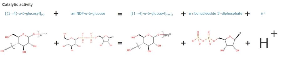
This is a reaction allowing the extension of amylose. It has a major role in the pathway starch biosynthesis.
[SSG1_ORYSJ] Required for the synthesis of amylose in endosperm.[HAMAP-Rule:MF_00484]
Publication Abstract from PubMed
The catalytic domain of rice (Oryza sativa japonica) granule bound starch synthase I (OsGBSSI-CD) was overexpressed and the three-dimensional structures of the ligand-free and ADP-bound forms were determined. The structures were similar to those reported for bacterial and archaeal glycogen synthases, which belong to glycosyltransferase family 5. They had Rossmann fold N- and C-domains connected by canonical two-hinge peptides, and an interdomain disulfide bond that appears to be conserved in the Poaceae plant family. The presence of three covalent linkages might explain why both OsGBSSI-CD structures adopted only the closed domain arrangement.
Interdomain Disulfide Bridge in the Rice Granule Bound Starch Synthase I Catalytic Domain as Elucidated by X-Ray Structure Analysis.,Momma M, Fujimoto Z Biosci Biotechnol Biochem. 2012 Aug 7. PMID:22878205[1]
From MEDLINE®/PubMed®, a database of the U.S. National Library of Medicine.
Disease
The protein 3vuf has allergenic properties, for example in mammals it can bind to Immunoglobulin E (IgE) causing an allergic response.
Relevance
Structure
Primary structure (amino acid sequence):
MSALTTSQLA TSATGFGIAD RSAPSSLLRH GFQGLKPRSP AGGDATSLSV
TTSARATPKQ QRSVQRGSRR FPSVVVYATG AGMNVVFVGA EMAPWSKTGG
LGDVLGGLPP AMAANGHRVM VISPRYDQYK DAWDTSVVAE IKVADRYERV
RFFHCYKRGV DRVFIDHPSF LEKVWGKTGE KIYGPDTGVD YKDNQMRFSL
LCQAALEAPR ILNLNNNPYF KGTYGEDVVF VCNDWHTGPL ASYLKNNYQP
NGIYRNAKVA FCIHNISYQG RFAFEDYPEL NLSERFRSSF DFIDGYDTPV
EGRKINWMKA GILEADRVLT VSPYYAEELI SGIARGCELD NIMRLTGITG
IVNGMDVSEW DPSKDKYITA KYDATTAIEA KALNKEALQA EAGLPVDRKI
PLIAFIGRLE EQKGPDVMAA AIPELMQEDV QIVLLGTGKK KFEKLLKSME
EKYPGKVRAV VKFNAPLAHL IMAGADVLAV PSRFEPCGLI QLQGMRYGTP
CACASTGGLV DTVIEGKTGF HMGRLSVDCK VVEPSDVKKV AATLKRAIKV
VGTPAYEEMV RNCMNQDLSW KGPAKNWENV LLGLGVAGSA PGIEGDEIAP
LAKENVAAP
Secondary structure:

Structural highlights
This protein is an enzyme, which is formed from one monomer and can bind to ADP. It binds to the ADP-glucose with amino acids 97,100, 408, 413,462 and 492. This allow the formation of an hydrophobic pocket in which ADP-glucose can binds to and in which he is protected from water and hydrolyse. we can also see that 3vuf can binds sulfate. This hydrophobic cage is allowed by the beta sheets and alpha helices that the protein have.
3vuf is a 1 chain structure with sequence from Japanese rice. Full crystallographic information is available from OCA. For a guided tour on the structure components use FirstGlance.
|
| Ligands: | , |
| Related: | 3vue |
| Gene: | WAXY, WX, WX-B, Os06g0133000, LOC_Os06g04200, 134P10.7, P0679C08.19 (Japanese rice) |
| Activity: | NDP-glucose--starch glucosyltransferase, with EC number 2.4.1.242 |
| Resources: | FirstGlance, OCA, PDBe, RCSB, PDBsum, ProSAT |
This is a sample scene created with SAT to by Group, and another to make of the protein. You can make your own scenes on SAT starting from scratch or loading and editing one of these sample scenes.


