Sandbox Reserved 1790
From Proteopedia
This page, as it appeared on June 14, 2016, was featured in this article in the journal Biochemistry and Molecular Biology Education.
Contents |
Lysophosphatidic Acid Receptor 1
Introduction
Lysophosphatidic Acid Receptor
Lysophosphatidic Acid
Structure
Structural Stabilization
Key Ligand Interactions

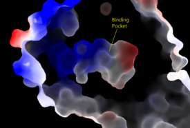
Figure 3: Electrostatic illustration of the amphipathic binding pocket of the LPA1 receptor. This binding pocket was revealed by cutting away the exterior or the protein. This binding pocket, located in the interior of the protein, has both polar and nonpolar regions. The blue and red coloration highlight the positively and negatively charged regions, respectively, and the white color shows the nonpolar region of the binding pocket.
Function
Receptor Comparison
Sphingosine 1-Phosphate Receptor
Endocannabinoid Receptor 1
Disease Relevance
Cancer
Pain
Fibrosis
3D structures of lysophosphatidic acid receptor
4z34, 4z35, 4z36 - hLPA1 + antagonist - human
2lq4 – hLPA1 second extracellular loop – NMR
4p0c – hLPA2/NHERF2
5xsz – LPA6A (mutant) – zebra fish
References
Proteopedia Resources
Category:Lysophosphatidic acid binding
Category:Lysophosphatidic acid
Butler University Proteopedia Pages
See also:
</StructureSection>
Student Contributors
Heather Hansen
Stephanie Kuhlman
Chandler Mitchell
Clayton Taylor


