Ubiquitin Structure & Function
From Proteopedia
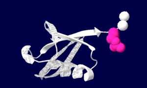
Ubiquitin is a single 8565 Mr polypeptide consisting of 76 amino acid residues. Ub is highly known for its role in ATP-dependant protein degradation.
| |||||||||
| 1ubq, resolution 1.80Å () | |||||||||
|---|---|---|---|---|---|---|---|---|---|
| |||||||||
| |||||||||
| Resources: | FirstGlance, OCA, PDBsum, RCSB | ||||||||
| Coordinates: | save as pdb, mmCIF, xml | ||||||||
Introduction
Ubiquitin is one of the most highly conserved eukaryotic proteins. Primary structures found throughout ubiquitin are identical in all bovine, insects and human ubiquitin. The only difference observed amongst these species is seen in the terminal Gly-Gly residues. Yeast and oat ubiquitin only differ in three of the 76 residues when compared to ubiquitin found in higher eukaryotes. Ubiquitin can not only be found in the nucleus, but in the cytoplasm and cell-surface membrane as well. One interesting characteristic of ubiquitin is its stability. Ubiquitin is able to withstand a range of pH levels and temperatures and is very resistant to tryptic digestion, while still containing seven Lysine and four arginine residues. Many aspects of ubiquitin's structure aid in this durability. Ubiquitin contains a hydrophobic core. Three hydrophobic residues found on the α-helix and 11 of the 13 hydrophobic residues from the β-sheet are involved in constructing this hydrophobic core. The main contributor to the ubiquitin stability is the vast amount of hydrogen-bonding interactions observed. The whole structure of ubiquitin undergoes significant hydrogen bonding, aside from the COOH terminus.
Function
At first, ubiquitin was believed to be a hormone involed in inducing the differentiation of lymphocytes and activating adenylate cyclase [1]. However, today ubiquitin is primarily known for its role in intracellular ATP-dependent protein degradation. This is accomplished by the formation of covalent conjugates between carboxyl terminals of ubiquitin and the target protein.
- ↑ Vijay-Kumar, S., Begg, CE., Wilkinson, KD and Cook, WJ. 1985. Three-dimensional structure of ubiquitin of 2.8 angstrom resolution. Proc Natl Acad Sci 82:3582-3585
Proteopedia Page Contributors and Editors (what is this?)
Jaclyn Gordon, Joel L. Sussman, Michal Harel, David Canner, Andrea Gorrell, Alexander Berchansky, Karsten Theis


