Sandbox Reserved 329
From Proteopedia
| This Sandbox is Reserved from January 10, 2010, through April 10, 2011 for use in BCMB 307-Proteins course taught by Andrea Gorrell at the University of Northern British Columbia, Prince George, BC, Canada. |
To get started:
More help: Help:Editing |
Contents |
Uridylyl transferases
INTRODUCTION
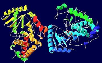
Terminal uridylyl transferases (TUTases) belong to a superfamily of polymerase ß nucleotidyl transferases.[1] TUTases have been isolated from Trypanosoma brucei and also Leishmania ssp, parasites that cause diseases in humans such as African Sleeping Sickness.[2] TUTases can function in RNA editing; more specifically TUTase4 catalyzes a reaction that adds a nucleotide, from a nucleotide triphosphate, to uridine monophosphate (UMP), the minimally required terminal RNA substrate. (ref) TUTase4 is able to bind to the nucleotide triphosphates ATP, CTP, GTP or UTP, however, UTP and CTP are preferred, whereas ATP and GTP ligands have been shown to cause a significant decrease in enzymatic activity (ref). The preference for UTP causes TUTase4 to typically add a uracil nucleotide to the RNA substrate. This selectivity has a variety of mechanisms, including a loss of coplanarity (pi-electron stacking) between the ATP and a tyrosine of the active site (Y189) required for catalysis, and reduced stacking between the UMP and ATP rings. The RNA substrate in trypanosomal TUTases selects for cognate nucleosides and provides a metal ion binding site for Mg2+ ions required by the ligand.[1]
| |||||||||
| 2q0d, resolution 2.00Å () | |||||||||
|---|---|---|---|---|---|---|---|---|---|
| Ligands: | , | ||||||||
| Gene: | TUT4 (Trypanosoma brucei) | ||||||||
| Activity: | RNA uridylyltransferase, with EC number 2.7.7.52 | ||||||||
| Related: | 2ikf, 2nom | ||||||||
| |||||||||
| |||||||||
| Resources: | FirstGlance, OCA, RCSB, PDBsum | ||||||||
| Coordinates: | save as pdb, mmCIF, xml | ||||||||
STRUCTURE
The uridylyl transferase bound ligand is an with two Mg2+ ions, however many TUTases involved in RNA editing are shown to exhibit preference for binding to UTP instead.[1] Three are conserved in TUTases, and are required for coordinating the Mg2+ ions in some TUTases. [1] Thus, these are vital in catalyzing this reaction.
REFERENCES
- ↑ 1.0 1.1 1.2 1.3 Stagno J, Aphasizheva I, Aphasizhev R, Luecke H. Dual role of the RNA substrate in selectivity and catalysis by terminal uridylyl transferases. Proc Natl Acad Sci U S A. 2007 Sep 11;104(37):14634-9. Epub 2007 Sep 4. PMID:17785418
- ↑ Aphasizhev R, Sbicego S, Peris M, Jang SH, Aphasizheva I, Simpson AM, Rivlin A, Simpson L. Trypanosome mitochondrial 3' terminal uridylyl transferase (TUTase): the key enzyme in U-insertion/deletion RNA editing. Cell. 2002 Mar 8;108(5):637-48. PMID:11893335


