From Proteopedia
proteopedia linkproteopedia link
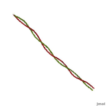
Tropomyosin (TPM) has a 4-helix coiled structure. It regulates the binding of myosin thus regulating muscle contraction [1]. In its locked conformation it binds troponin T (TnnT) and prevents the binding of myosin to actin. When Ca++ ions bind to TnnT, the TPM assumes an open conformation and myosin can bind to actin. The images on the left and the right correspond to one representative TPM structure, i.e. tropomyosin from pig (1c1g). You can at the right for clarity. The dimers of TPM in the asymmetric unit (1c1g) are , with their C-terminal ends overlapping by about 2/3 of the molecular length. This suggests head-to-tail packing of TPM, which is very important for its interaction with actin [2].
3D Structures of Tropomyosin
3mtu, 3mud – cTPM alpha-1 – chicken
1ic2 - cTPM alpha-1 (mutant)
2w49, 2w4u – cTnnC+cTnnT+cTnnI+cTPM alpha-1+cActin
2z5h – yTPM alpha-1 N-terminal+C-terminal+GNC4 leucine zipper+TnnT – yeast
2z5i - yTPM alpha-1 N-terminal+C-terminal+GNC4 leucine zipper
2efr, 2efs, 2d3e - rTPM alpha-1 C-terminal+GNC4 leucine zipper – rabbit
1kql - TPM alpha-1 C-terminal+GNC4 leucine zipper - rat
1mv4 - TPM alpha-1 C-terminal – rat
2g9j - TPM alpha-1 TM9A+GNC4 – rat
2b9c – TPM mid region – rat
1c1g – TPM – pig
2tma – TPM - model
References
- ↑ Gunning P, Weinberger R, Jeffrey P. Actin and tropomyosin isoforms in morphogenesis. Anat Embryol (Berl). 1997 Apr;195(4):311-5. PMID:9108196
- ↑ Whitby FG, Phillips GN Jr. Crystal structure of tropomyosin at 7 Angstroms resolution. Proteins. 2000 Jan 1;38(1):49-59. PMID:10651038

