Sandbox Reserved 481
From Proteopedia
| This Sandbox is Reserved from 13/03/2012, through 01/06/2012 for use in the course "Proteins and Molecular Mechanisms" taught by Robert B. Rose at the North Carolina State University, Raleigh, NC USA. This reservation includes Sandbox Reserved 451 through Sandbox Reserved 500. | |||||||
To get started:
More help: Help:Editing For more help, look at this link: http://www.proteopedia.org/wiki/index.php/Help:Getting_Started_in_Proteopedia
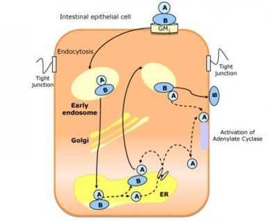
Cholix Toxin from Vibrio choleraeIntroductionCholix toxin (CT) are a class of protein toxin originating from the bacteria Vibrio cholerae. It is the third memeber of eEF2-specific ADP-ribosyltransferase toxins. The toxin uses ADP-riboyltransferase to inactivate eukaryotic elongation factor 2 by transferring ADP-ribose from NAD+ which inhibits protein synthesis and causes cell death. This protein toxin has been known to cause disease in both plants and animals. Specifically, the toxin can cause the disease Cholera. It enters eukaryotic cells through endocytosis. Once inside, the toxin transfers an ADP-ribose group to an Arg residue of the GTP binding protein G. This then activates adenylate cyclase which leads to an increase amount of cAMP, causing a secretion of Cl-,HCO3-, and water from epithelial cells from the site of colonization. The result is dehydration and loss of electrolytes in infected hosts. Cholix toxins are composed of a receptor binding, translocation, and catalytic domain. StructureX-Ray CrystallographyTo observe the structure, the Cholix Toxin was crystallized by vapor diffusion against reservoirs containing 23% polyethlene gylcol-10,000, 7.5% ethylene glycol, and 0.1 m HERPES. In addition, the reservoirs were kept at a of pH 7.5 and at a temperature of 19°C. About 40 µL of reservior solution containg 1.25 mM NAD+ solution was added to a 2 µL crystal containing group. The NAD+ was allowed to soak into the crystals for approximately 2-3 minutes. The crystals were then transferred to paratone-N for visualization via X-Ray Diffraction Protein StructureThe sequence of the Cholix Toxin is 666 amino acids in length. It contains a total of four disulfide bonds which are located between Cys-43:Cys-47, Cys-240:Cys: 257, Cys-310:Cys-332, and Cys-426:Cys-433. The secondary structure of the Cholix Toxin consists of 11 and 4 The Cholix Toxin contains three different domains. Though it is not shown, the active site for this toxin is located at Glu-613. MoleculeThe Cholix Toxin is a bipartite molecule consisting of 1 A subunit which further divides into two peptides: A1 and A2 and 5 B subunits. The A subunit is considered the active subunit while the B subunit serves to bind to GM1 ganglioside on the surfaces of intestinal epithelial cells. Both of these subunits are highly essential for the functionality of this toxin.
Mechanism of ActionBasic MechanismThe mechanism for the cholix toxin occurs as follows:
ADP-ribosylation of Gs-α subunit of G-ProteinsDiseasesReferences1. The 1.8A Cholix Toxin Crystal Structure in Complex with NAD+ and Evidence for a New Kinetic Model |

