1vgj
From Proteopedia
|
Crystal structure of 2'-5' RNA ligase from Pyrococcus horikoshii
Overview
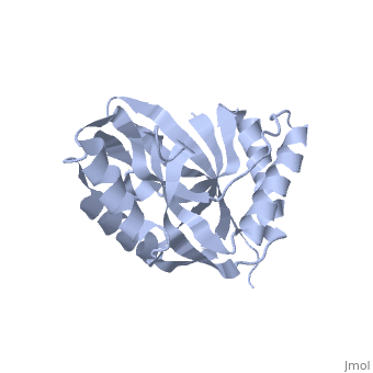
Bacterial and archaeal 2'-5' RNA ligases, members of the 2H, phosphoesterase superfamily, catalyze the linkage of the 5' and 3' exons, via a 2'-5'-phosphodiester bond during tRNA-precursor splicing. The, crystal structure of the 2'-5' RNA ligase PH0099 from Pyrococcus, horikoshii OT3 was solved at 1.94 A resolution (PDB code 1vgj). The, molecule has a bilobal alpha+beta arrangement with two antiparallel, beta-sheets constituting a V-shaped active-site cleft, as found in other, members of the 2H phosphoesterase superfamily. The present structure was, significantly different from that determined previously at 2.4 A, resolution (PDB code 1vdx) in the active-site cleft; the entrance to the, cleft is wider and the active site is easily accessible to the substrate, (RNA precursor) in our structure. Structural comparison with the 2'-5' RNA, ligase from Thermus thermophilus HB8 also revealed differences in the RNA, precursor-binding region. The structural differences in the active-site, residues (tetrapeptide motifs H-X-T/S-X) between the members of the 2H, phosphoesterase superfamily are discussed.
About this Structure
1VGJ is a Single protein structure of sequence from Pyrococcus horikoshii. Full crystallographic information is available from OCA.
Reference
The structure of Pyrococcus horikoshii 2'-5' RNA ligase at 1.94 A resolution reveals a possible open form with a wider active-site cleft., Gao YG, Yao M, Okada A, Tanaka I, Acta Crystallograph Sect F Struct Biol Cryst Commun. 2006 Dec 1;62(Pt, 12):1196-200. Epub 2006 Nov 30. PMID:17142895
Page seeded by OCA on Wed Nov 21 04:49:27 2007
