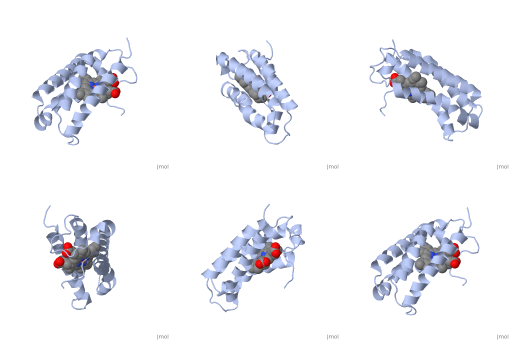
1a7v
From Proteopedia
|
CYTOCHROME C' FROM RHODOPSEUDOMONAS PALUSTRIS
Overview
The crystal structure of an unusual monomeric cytochrome c' from Rhodopseudomonas palustris (RPCP) has been determined at 2.3 A resolution. RPCP has the four-helix (helices A, B, C and D) bundle structure similar to dimeric cytochromes c'. However the amino acid composition of the surface of helices A and B in RPCP is remarkably different from that of the dimeric cytochromes c'. This surface forms the dimer interface in the latter proteins. RPCP has seven charged residues on this surface contrary to the dimeric cytochromes c', which have only two or three charged groups on the corresponding surface. Moreover, hydrophobic residues on this surface of RPCP are two to three times fewer than in dimeric cytochromes c'. As a result of the difference in amino acid composition, the A-B surface of RPCP is rather hydrophilic compared with dimeric cytochromes c'. We thus suggest that RPCP is monomeric in solution because of the hydrophilic nature of the A-B surface. The amino acid composition of the A-B surface is similar to that of Rhodobacter capsulatus cytochrome c' (RCCP), which is an equilibrium admixture of monomer and dimer. The charge distribution of the A-B surface in RCCP, however, is considerably different from that of RPCP. Due to the difference, RCCP can form dimers by both ionic and hydrophobic interactions. These dimers are quite different from those in proteins which form strong dimers such as in Chromatium vinosum, Rhodospirillum rubrum, Rhodospirillum molischianum and Alcaligenes. Cytochrome c' can be classified into two types. Type 1 cytochromes c' have hydrophobic A-B surfaces and they are globular. The A-B surface of type 2 cytochromes c' is hydrophilic and they take a monomeric or flattened dimeric form.
About this Structure
1A7V is a Single protein structure of sequence from Rhodopseudomonas palustris with as ligand. Full crystallographic information is available from OCA.
Reference
Basis for monomer stabilization in Rhodopseudomonas palustris cytochrome c' derived from the crystal structure., Shibata N, Iba S, Misaki S, Meyer TE, Bartsch RG, Cusanovich MA, Morimoto Y, Higuchi Y, Yasuoka N, J Mol Biol. 1998 Dec 4;284(3):751-60. PMID:9826513
Page seeded by OCA on Thu Feb 21 11:41:50 2008

