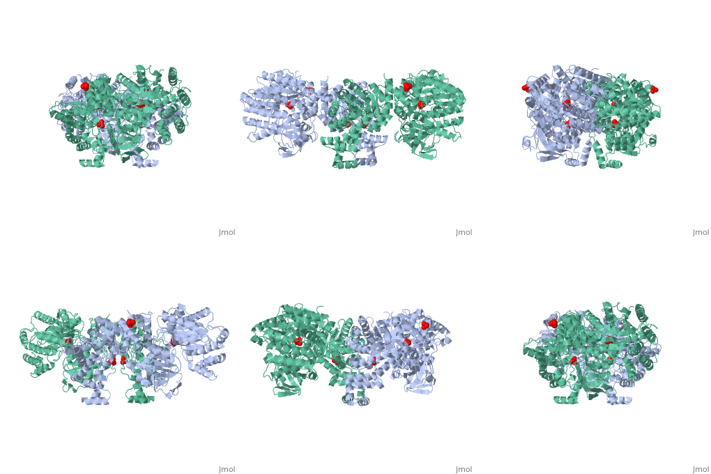
1dik
From Proteopedia
|
PYRUVATE PHOSPHATE DIKINASE
Overview
The crystal structure of pyruvate phosphate dikinase, a histidyl multiphosphotransfer enzyme that synthesizes adenosine triphosphate, reveals a three-domain molecule in which the phosphohistidine domain is flanked by the nucleotide and the phosphoenolpyruvate/pyruvate domains, with the two substrate binding sites approximately 45 angstroms apart. The modes of substrate binding have been deduced by analogy to D-Ala-D-Ala ligase and to pyruvate kinase. Coupling between the two remote active sites is facilitated by two conformational states of the phosphohistidine domain. While the crystal structure represents the state of interaction with the nucleotide, the second state is achieved by swiveling around two flexible peptide linkers. This dramatic conformational transition brings the phosphocarrier residue in close proximity to phosphoenolpyruvate/pyruvate. The swiveling-domain paradigm provides an effective mechanism for communication in complex multidomain/multiactive site proteins.
About this Structure
1DIK is a Single protein structure of sequence from Clostridium symbiosum with as ligand. Active as Pyruvate, phosphate dikinase, with EC number 2.7.9.1 Full crystallographic information is available from OCA.
Reference
Swiveling-domain mechanism for enzymatic phosphotransfer between remote reaction sites., Herzberg O, Chen CC, Kapadia G, McGuire M, Carroll LJ, Noh SJ, Dunaway-Mariano D, Proc Natl Acad Sci U S A. 1996 Apr 2;93(7):2652-7. PMID:8610096
Page seeded by OCA on Thu Feb 21 12:16:56 2008

