2pea
From Proteopedia
| |||||||||
| 2pea, 1 NMR models () | |||||||||
|---|---|---|---|---|---|---|---|---|---|
| Related: | 1d3z, 1aar, 2bgf, 2pe9 | ||||||||
| |||||||||
| |||||||||
| Resources: | FirstGlance, OCA, PDBsum, RCSB | ||||||||
| Coordinates: | save as pdb, mmCIF, xml | ||||||||

Contents |
NMR Based Structure of the Closed Conformation of LYS48-Linked Di-Ubiquitin Using Experimental Global Rotational Diffusion Tensor from NMR Relaxation Measurements
Template:ABSTRACT PUBMED 17550252
About this Structure
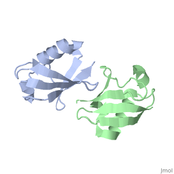
2pea is a 2 chain structure with sequence from Homo sapiens. Full experimental information is available from OCA.
See Also
Reference
- Ryabov Y, Fushman D. Structural assembly of multidomain proteins and protein complexes guided by the overall rotational diffusion tensor. J Am Chem Soc. 2007 Jun 27;129(25):7894-902. Epub 2007 Jun 6. PMID:17550252 doi:10.1021/ja071185d
- Ryabov YE, Geraghty C, Varshney A, Fushman D. An efficient computational method for predicting rotational diffusion tensors of globular proteins using an ellipsoid representation. J Am Chem Soc. 2006 Dec 6;128(48):15432-44. PMID:17132010 doi:10.1021/ja062715t
- Ryabov YE, Fushman D. A model of interdomain mobility in a multidomain protein. J Am Chem Soc. 2007 Mar 21;129(11):3315-27. Epub 2007 Feb 24. PMID:17319663 doi:10.1021/ja067667r
- Varadan R, Walker O, Pickart C, Fushman D. Structural properties of polyubiquitin chains in solution. J Mol Biol. 2002 Dec 6;324(4):637-47. PMID:12460567
