This is a default text for your page Ivan Koutsopatriy estrogen receptor. Click above on edit this page to modify. Be careful with the < and > signs.
You may include any references to papers as in: the use of JSmol in Proteopedia [1] or to the article describing Jmol [2] to the rescue.
There are two flavors of estrogen receptors. ER - Intracellular receptor and GPER – G protein coupled receptors. ERs are transcription factors while GPER are not transcription factors. ERs are the focus of this page.
Function
Like other steroid hormones estrogen effects the transcription of a large number of genes via its interactions with its intracellular receptors, estrogen receptors. Estrogen receptors are receptors that are activated by the hormone estrogen (Example of an estrogen; 17β-estradiol).
Disease
Relevance
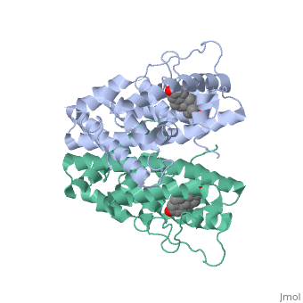
Structural highlights
ER is a modular protein composed of a ligand binding domain, a DNA binding domain and a transactivation domain. ER is a DNA-binding transcription factor. Unbound ER normally exists loosly around the nucleus; this is subject to change depending on a multitude of factors including cell type, progress through cell cycle and reception of cellular signals. When estrogen enters the cell and binds ER, ER will trans-locates and undergoes a conformational shift.(1) Ligand bound estrogen receptor associates more tightly with the nucleus.
The specific conformation of this tight loop creates part of the activation signal that will stimulate normal growth, as estradiol is a normal ligand for ER.
Tamoxifen is a drug created to bind ER and inhibit the transcription factor activity of ER. Tamoxifen is larger than the normal hormone ER binds (estradiol); for this reason the activation loop is pushed into an inactive conformation. This blocks ER from giving the signal to grow.
ERα-regulated gene expression involves interactions with cointegrators (ex. p300/CBP, P/CAF) that have the capacity to modify core histone acetyl groups. 2 ER’s DNA binding domain is ordered around two zinc ions that allow the receptors to bind as homodimers to palindromic DNA sequences in such a way that each homodimer links to one half of the palindrome.
This is a sample scene created with SAT to by Group, and another to make of the protein. You can make your own scenes on SAT starting from scratch or loading and editing one of these sample scenes.