Sanbox glut3
From Proteopedia
Facilitated Glucose Transporter 3, Solute Carrier Family 2 (GLUT3/ SLC2A3) in Homo Sapiens
This is a default text for your page Sanbox glut3. Click above on edit this page to modify. Be careful with the < and > signs. You may include any references to papers as in: the use of JSmol in Proteopedia [1] or to the article describing Jmol [2] to the rescue.
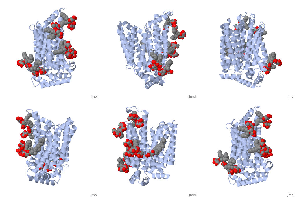
Function is one of fourteen facilitative sugar transporters which use the glucose diffusion gradient to move across various plasma membranes to display various specificities, kinetics and tissue expression profilesCite error: Invalid Structure, Disease in HumansAlzheimer’s disease, shows levels of impaired glucose uptake and metabolism, which leads to the downgrade of many other factors in the brain. GLUT3 is responsible for transporting glucose from extracellular space to neuronal tissue (i.e. dendrites and axons). Decreased levels of GLUT3 in Alzheimer brain shows a positive correlation to decreased levels of N-acetylglucosamine. The impaired presence of GLUT3 leads to hyperphosphorylation of the Tau protein, which normally stabilizes neuronal microtubules. Lastly there is a reduction in the transcription for factor hypoxia-inducible factor 1, which plays a role in glucose metabolism in the brain. The comparison between a normal healthy brain and an Alzheimer brain relieved that there was a 25-30% decrease in GLUT3 levels in the Alzheimer brain. Possible MechanismMultiple mechanistic theories have been proposed for facilitated glucose transporters. The simple carrier model was the earliest theory proposed by Widdas and contains four steps. First, the empty carrier opens to the cis side of the membrane for glucose to bind1. Then the substrate binding carrier translocates to the trans side of the membrane where it then releases glucose on that side. Last the empty carrier switches to the cis side. Multiple mechanistic theories, including the simple carrier model were proposed but all attempted to explain two key components of GLUT transporters, the asymmetry of the transport affinities and the trans-acceleration that occurs in the presence of hexose on the trans side2. After considerable research, two popular models remain for class 1 glut transporters. The two-site/fixed site transporter theory explains the asymmetry by having both substrate binding sites simultaneously available3. After glucose is bound, hexoses exchange between sites and speed the binding process. Although this method explains the asymmetry and the kinetics of class 1 glut transporters, not all class 1 glut transporters contain asymmetric glucose transporters3. The alternating access model follows three steps4. The transporter has a cavity for small substrates, and contains a substrate binding site. The transporter also has two different configurational openings to one cell membrane or the other. This mechanisms differs from the two-site/fixed site transporter theory by assuming there is only one binding site available at a time, leading to four different conformation states. An empty outward open state, an occluded transporter state, a inward open state and finally another occluded state5. Trans-acceleration is only observed in a minority of class 1 glut transporters6. GLUT3 has been proven to be dependent on trans-acceleration. This method was discovered when hexose was found to be moving against its concentration gradient1. This movement is argued to support both the two-site transporter theory and the alternating access model. Geminate exchange, named by Naftalin et al, explains this movement with the idea that hexose could exchange freely between two binding sites within the carrier. While other scientist argue that hexose could move from outward to inward without glucose binding7. RelevanceStructural highlightsThis is a sample scene created with SAT to by Group, and another to make of the protein. You can make your own scenes on SAT starting from scratch or loading and editing one of these sample scenes.
| ||||||||||||
References
- ↑ Hanson, R. M., Prilusky, J., Renjian, Z., Nakane, T. and Sussman, J. L. (2013), JSmol and the Next-Generation Web-Based Representation of 3D Molecular Structure as Applied to Proteopedia. Isr. J. Chem., 53:207-216. doi:http://dx.doi.org/10.1002/ijch.201300024
- ↑ Herraez A. Biomolecules in the computer: Jmol to the rescue. Biochem Mol Biol Educ. 2006 Jul;34(4):255-61. doi: 10.1002/bmb.2006.494034042644. PMID:21638687 doi:10.1002/bmb.2006.494034042644

