Introduction
Every bacteria wants to live. Every bacteria wants to reproduce. To achieve both, bacteria need some sort of protection that will maintain the structure inside and will protect it from outside dangers. This protection is called cell wall, that primarily consists of a polymer that is called peptidoglycan. This compound can be synthesized only with the help of penicillin binding proteins (PBPs) , which are the target of this article (the name basically comes from a way it was discovered).
The goal of a chemist is to develop a way to break this cell wall, thus destroying the bacteria. To achieve this, the penicillin is introduced to the bacteria and then reacts with PBPs (using β-lactam ring ), preventing it from forming a cell wall. However, many pathogenic bacteria have evolved a way to mutate themselves to be immune to various drugs that contain this β-lactam rings. What happens is that bacterium produces enzyme, called that cleaves the β-lactam ring on a penicillin and thus preventing it from reacting with PBPs. To solve this problem, the new drug, called, lactivicin was developed that contains gamma-lactone rings and cycloserine as substitutions to β-lactam. So far, it has proved to be an efficient antibiotic. It successfully binds to PBPs and prevents cell wall from forming.
Recently, an analog of lactivicin, phenoxylactivicin (PLTV) was developed and is discussed in this article.
The complex of the PBP with is shown on the picture.
Picture on the right is displayed as N-terminus to C-termiunus Rainbow for. The coloring goes as shown on the sample:
| Amino Terminus |
|
|
|
|
|
|
|
Carboxy Terminus |
Overall Structure
Penicillin Binding Proteins have specific structures and designs that promote allow the binding of Penicillin and other antibiotics. One of the enzymes within the PBP family is D-alanyl-D-alanine carboxypeptidase/transpeptidase. This enzyme is responsible for the link between two chains in the peptidoglycan network [1]. DA-DA peptidase’s structure contains a serine in the active site. Ser 62 is used to bind a peptide strand which would then link to another strand of the network, and this is the site where penicillin binds and inhibits the protein.
This enzyme is split into two sections, which will be referred to as the North and South regions. The North Region contains both the carboxyl and amino termini, two α-helices, and a nine-stranded antiparallel β-sheet [2]. This leads the Northern region of the enzyme to appear symmetrical. Both termini lead are connected to helices and then into β-strands. Inbetween the sets of strands the South region of the peptide is formed and this is strictly made out of helices. In the center of the two regions is where the Ser 62 active site resides, and this is also at the symmetrical center of the protein. The protein essentially forms a cupped hand, with the center of the palm being the active site, the bottom of the palm being a series of 8 or so helices, the knuckles being the β-strands, and the tips of the fingers being the two helices of the North region.
Binding Interactions
Penicillin binding protein binds to beta-lactam antibiotics because they are similar in chemical structure to the modular pieces that form the peptioglycan
. The amide bond
is ruptured to form a covalent bond with the catalytic serine at the binding protein's active site.
Additional Features
1)role in antibiotics and resistance
the role of PBPs in its synthesis is a very good target for drugs of selective toxicity
2)different PBPs and how they are classified into diverse categories
found as both membrane-bound and cytoplasmic proteins
3)work as enzyme to catalyze synthesis of peptidoglycan
Quiz Question 1
Question related to PBP evolution and developing resistance to ß-lactams, likely related to active site mutations.
Shows the from most conserved to most variable.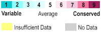
See Also
Credits
Introduction - Anton El Khoury
Overall Structure - Tyler Carpenter
Drug Binding Site - Hyunjoon Choi
Additional Features - Tiankai Zhang
Quiz Question 1 - Samuel Pierce
References
- ↑ Macheboeuf P, Fischer DS, Brown T Jr, Zervosen A, Luxen A, Joris B, Dessen A, Schofield CJ. Structural and mechanistic basis of penicillin-binding protein inhibition by lactivicins. Nat Chem Biol. 2007 Sep;3(9):565-9. Epub 2007 Aug 5. PMID:17676039 doi:10.1038/nchembio.2007.21
1. Goodsell, David. "Penicillin-binding Proteins." Penicillin-binding Proteins. May 2002. Web. 07 Apr. 2016.
2. Kelly, J. A., and A. P. Kuzin. "3PTE." RCSB PDB. Web. 07 Apr. 2016.

