Sandbox Reserved 1063
From Proteopedia
Adhesin Competence Regulator
Introduction

Adhesin Competence Regulator (AdcR) is a transcriptional regulator that controls the activation of over seventy genes within the bacteria Streptococcus pneumoniae[1]. Contrasting with other members of the multiple antibiotic resistance regulator (MarR) family, AdcR is metal dependent as the binding of Zinc causes conformational changes that permit AdcR to repress the transcription of high-affinity Zinc specific uptake transporters. Zinc plays a vital role in organism homeostasis acting as a co-factor and a regulator of enzymatic activity, however zinc can lead to cell toxicity and deficiency of other vital metals that are also necessary for protein function[2][3]. Given the many roles zinc plays in general homeostasis the importance of AdcR in Streptococcus pneumoniae can be understood provided its ability to regulate zinc transfer proteins within the bacteria.

Members of the MarR protein family conserve a number of features including a general triangular shape, a two fold pseudosymmetric homo dimer, and a wingled helix-turn-helix pattern. Consistent with AdcR's identity as a member of the MarR protein family, AdcR exhibits the triangular shape with the (wHTH) binding domain. This structure calls for multiple zinc binding sites that facilitate protein conformational change allowing for DNA binding and regulation through the wHTH domain.
| |||||||||||
References
- ↑ Sanson M, Makthal N, Flores AR, Olsen RJ, Musser JM, Kumaraswami M. Adhesin competence repressor (AdcR) from Streptococcus pyogenes controls adaptive responses to zinc limitation and contributes to virulence. Nucleic Acids Res. 2015 Jan;43(1):418-32. doi: 10.1093/nar/gku1304. Epub 2014 Dec, 15. PMID:25510500 doi:http://dx.doi.org/10.1093/nar/gku1304
- ↑ Fraústo da Silva J, Williams R. The Biological Chemistry of Elements: The Inorganic Chemistry of Life. Second ed. Oxford University Press; Oxford: 2001.
- ↑ Ma Z, Jacobsen FE, Giedroc DP. Coordination chemistry of bacterial metal transport and sensing. Chem Rev. 2009 Oct;109(10):4644-81. doi: 10.1021/cr900077w. PMID:19788177 doi:http://dx.doi.org/10.1021/cr900077w
- ↑ Guerra AJ, Dann CE, Giedroc DP. Crystal Structure of the Zinc-Dependent MarR Family Transcriptional Regulator AdcR in the Zn(II)-Bound State. J Am Chem Soc. 2011 Nov 21. PMID:22085181 doi:10.1021/ja2080532
- ↑ PMID:20804771<ref/ref>. This is due to the charges on the histidines of the binding site. At pH 8, the histidines are positively charged and can interact with the negatively charged Zn(II) ion. However, at pH 6 the histidines are neutrally charged and will not coordinate as well with Zn(II).
Contents
Binding Site 2
consists of a highly distorted tetrahedral geometry around the zinc ion. There are three amino acids involved in the binding of the zinc ion (C30, E41, and E107) as well as a water molecule. If a C30A AdcR missense is present in binding site 2, it will have no effect on the ability of the protein to bind DNA. Therefore, binding site 2 has no significant role in DNA binding.
Other Ligands
The AdcR MarR transcriptional regulator is able to bind Co(II) in binding site 1 in a way that induces similar conformational changes to Zn(II) binding. Co(II) coordination in binding site 1 is able to allosterically activate DNA binding similarly to Zn(II) binding.
DNA Binding
Hydrogen Bond Network
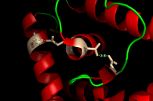 The Hydrogen Bonding Network is shown with dotted green lines approximately 2.8 angstroms between residues. The network consists of 4 major residues as follows from right to left: E24, N38, Q40, S74.
The binding of Zinc allows for the conformational change that induces the binding of DNA in order to activate genes. The binding of Zinc metals creates a hydrogen bond network within the protein that connects the metal binding sites and the DNA binding domain. More importantly, the hydrogen bonding network connects the metal binding pockets to the alpha 4 helix. Alpha 4 helix on each monomer plays a crucial role in binding DNA because it acts as the recognition helix. in the recognition helix recognize a sequence of DNA that is unknown at the moment; however, scientists do know that the hydrogen bond network acts as an allosteric activator for the protein to bind DNA. The hydrogen bond network connects the alpha 2 and alpha 4 helix via hydrogen bonding between specific residues. After zinc is bound, a glutamate (E24) residue from a random coil accepts a hydrogen bond from the carboxamide end of an asparagine (N38) residue from the alpha 2 helix. Then, a glutamine (Q40) residue from alpha 2 helix accepts a hydrogen bond from a serine (S74) residue from the alpha 4 helix. The color coding in the previous sentence represents the (), which is seen across the MarR family as a whole.
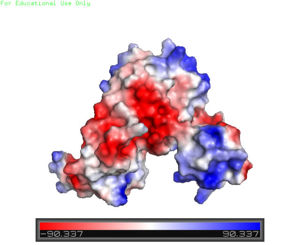 A charge map of AdcR shows the general triangular shape and the positive charged (blue) area on HTH domains
Helix-Turn-Helix Domain
The AdcR MarR transcriptional regulator's structure resembles the other proteins in the same family as mentioned before; however, the most notable differences are found in the winged helix-turn-helix (wHTH) motif that assists in binding DNA. The major groove of DNA is bound to the recognition helix while the wings grip onto the minor grooves of DNA. The charge map on the right highlights the positively charged areas, which stabilize the negatively charged backbone of DNA. Although AdcR is a highly alpha helical protein, the "wings" of the DNA binding domain consist of two anti parallel beta strands that are made up of several positively charged residues <ref>PMID:22085181</li></ol></ref>


