Sandbox Reserved 1239
From Proteopedia
| This Sandbox is Reserved from Jan 17 through June 31, 2017 for use in the course Biochemistry II taught by Jason Telford at the Maryville University, St. Louis, USA. This reservation includes Sandbox Reserved 1225 through Sandbox Reserved 1244. |
To get started:
More help: Help:Editing |
Contents |
Function and Location
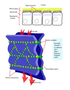
Occludin is one of the proteins absolutely necessary in maintaining tight junctions of cells. Tight junctions preserve the integrity of polarized cells, allowing homeostasis to continue. Polarized cells can be broadly defined as cells that have two dynamically different faces for functionally different purposes. Each face is different in protein and membrane composition. For example, intestinal cells have an apical surface and basolateral surface. The apical surface faces the lumen and intakes material, while the basolateral surface interacts with blood vessels. Proteins and lipids can diffuse laterally and freely through a membrane [1]. However, in cells that are meant to be polarized, it would be catastrophic to have proteins exchanged between apical and basolateral surfaces due to their pivotal roles in homeostasis. Tight junctions restrict lateral diffusion, keeping cell surfaces polarized [2]. Tight junctions, and therefore occluding proteins, heavily influence cell-cell interactions and signal transduction. Cell-cell interactions regulate gene expression and cell differentiation [5].
Structure
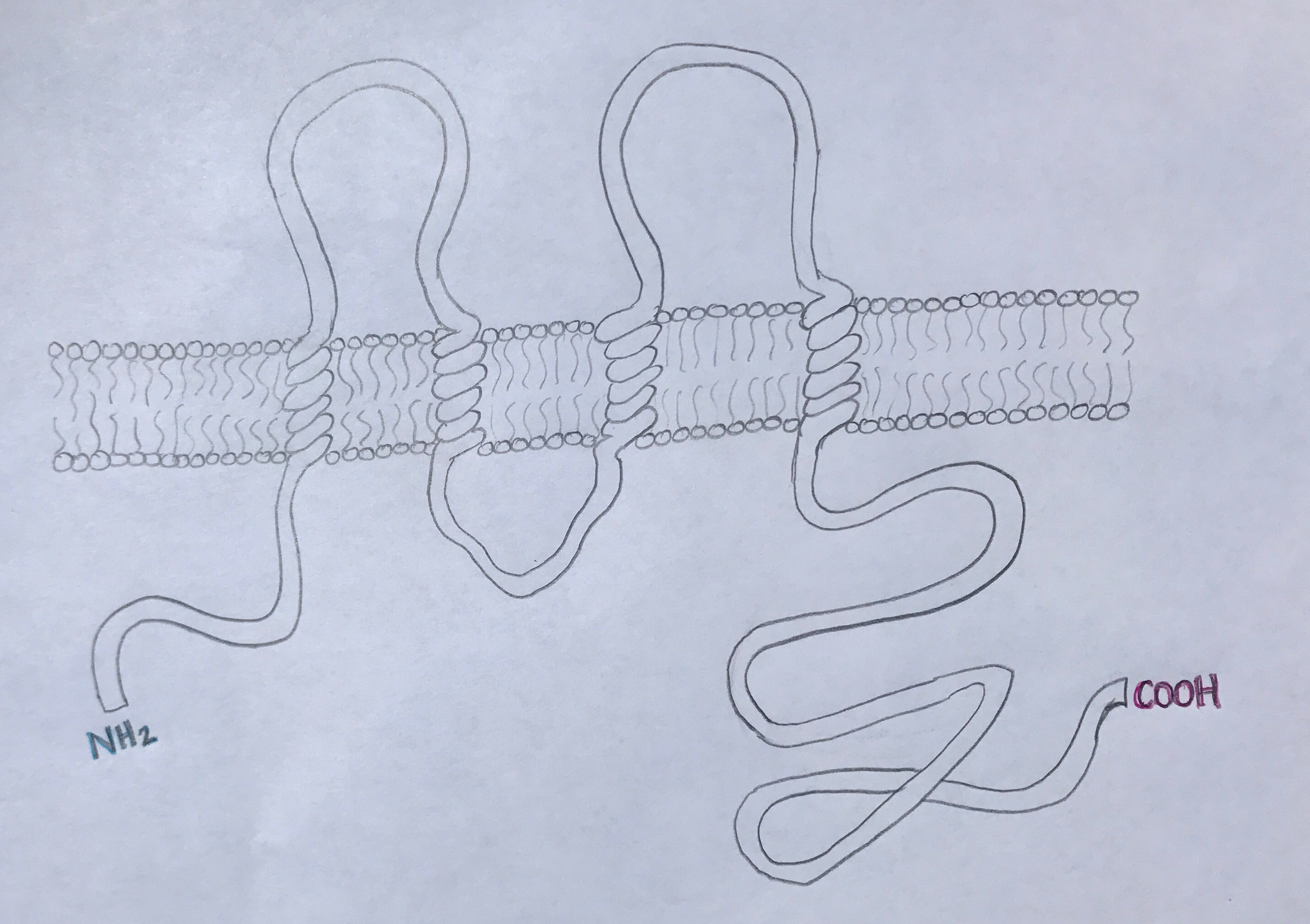
Occludin is approximately 65kDa in length. It was first identified in avian tissue, and was the first integral membrane tight junction protein to be found. Studies have found that occludin structure has 90% homology among mammals [3]. Occludin has four transmembrane alpha-helices, which are responsible for escorting occluding to tight junctions between cells [2]. As can be viewed in the image below, both the amino terminus and carboxyl terminus are located within the cytosol. The C-terminus of occluding is believed to interact with tight junction protein-1 (ZO-1), which is involved in cell-cell signaling [4].
Disease
Relevance
Structural highlights
This is a sample scene created with SAT to by Group, and another to make of the protein. You can make your own scenes on SAT starting from scratch or loading and editing one of these sample scenes.
</StructureSection>


