This old version of Proteopedia is provided for student assignments while the new version is undergoing repairs. Content and edits done in this old version of Proteopedia after March 1, 2026 will eventually be lost when it is retired in about June of 2026.
Apply for new accounts at the new Proteopedia. Your logins will work in both the old and new versions.
User:Jamie Abbott/Sandbox2
From Proteopedia
Contents |
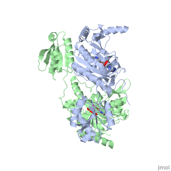
Histidyl-tRNA Synthetase
Histidyl tRNA Synthetase (HisRS) is a 94kD that belongs to the class II of aminoacyl-tRNA synthetases (aaRS). Aminoacyl-tRNA synthetases play a key role in protein synthesis and are classified as ligases. Aminoacyl tRNA synthetases use high energy ATP to attach a specific amino acid to its cognate tRNA[1]. This family of enzymes have been partitioned into two classes, containing 10 members, on the basis of sequence comparisons[1]. Class I and class II enzymes differ mainly with respect to the topology of the catalytic fold and site of esterification on cognate tRNA[1]. Class I aaRS enzymes contain a conserved Rossmann fold catalytic domain and often monomeric. Furthermore, class I enzymes attach the activated amino acid to the 2'OH of the tRNA molecule during catalysis, which then migrates to the 3'OH. Class II enzymes have a composed of anti-parallel (residues 1-325). Additionally, class II enzymes can be further divided into three subgroups: class IIa, distinguished by an N-terminal catalytic domain and C-terminal accessory domain (later shown to be the ); class IIb, whose anticodon binding domain is located in the N-terminal region; and class IIc, encompassing the tetrameric PheRS and GlyRS class II synthetases [2].
Histidyl-tRNA synthetases catalyze the transfer of histidine to a histidinyl transfer RNA molecule (tRNAHis). As HisRS is a class II aaRS enzyme it attaches the amino acid histidine to the 3’OH of the terminal ribose of tRNA[3]. Histidine is often an important amino acid in the active site of other enzymes and is unique as it can behave as either an acid or a base. The overall secondary structure of a HisRS monomer consist of 20% beta sheets and 37% helical character. It is structurally classified by CATH as an α-β layered sandwich. The CATH hierarchy of structural classification is based on: Class, Architecture, Topology, Homologous superfamily. Each of HisRS contains a N-terminal catalytic domain, C-terminal anticodon binding domain, a , II, and III, an insertion domain, a HisA and HisB loop, as well as an insertion domain. These structural elements are essential in assisting the two step mechanism carried out to aminoacylate tRNAHis with high fidelity.
Substrate Specificity
| |||||||||||

Mechanism of the Adenylation Reaction
 |  |  |
|
Electrophilic Catalysis
The HisRS active site contains a highly conserved residue in the HisRS family, Arg259, that takes part in electrophilic catalysis for the adenylation reaction. This active site arginine residue is not present in other aaRS class II enzymes. As mentioned previously are positioned to interact with the α-phosphate of ATP to assist in the adenylation reaction. Arg259 is positioned on the HisA loop to fix the α-carboxylate group of the histidine substrate as the [8]. Also, one ηN of the guanidinium group of Arg259 is positioned approximately 3Å from the α-phosphate of ATP while the other ηN hydrogen bonds with phenolic group of Tyr264. Thus, stabilization of the Tyr264 residue further enhances substrate binding allowing for the formation of a hydrogen bond to the Nδ of the histidine[3].
A comparison of in the HisRS:histidinol and the HisRS:adenylate complexes provides further structural information into how Arg259 may serve a role in catalysis. In the HisRS:histidinol complex a water-mediated interaction exists between Glu270 and εN of Arg259. However, in the HisRS:adenylate complex Glu270 moves to form a salt bridge with the guanidinium group excluding the water molecule. This movement serves as a salt bridge switch that may weaken the ionic interaction between Arg259 and the α-phosphate[4] and stabilize adenylate formation in the active site. Also, Arg113 as well as Arg259 are arranged to interact with of the histidyl-adenylate intermediate and stabilizes negative charge developed on the non-bridging oxygens α-phosphate during the transition state [3]. Evidence for Arg259 as critical residue in catalysis is further supported by mutational studies where a two or three log decrease in activity is observed when Arg259 is substituted with histidine [4] or other amino acids[9]. Utilizing Arg259 for catalysis is unique to HisRS as other class II aaRS enzymes, AspRS[10] and SerRS[7], use a divalent magnesium metal ion to coordinate the α-phosphate of ATP and serve as an electrophilic catalyst.
Mechanism of the Aminoacylation Reaction
 |  |  |

Substrate Assisted Catalysis
The second reaction carried out by HisRS, , requires the decomposition of a mixed anhydride (the ) to form an aminoacyl ester on the 3’OH of tRNAHis. It was initially hypothesized that Glu83, acting as a general base, would improve the rate of this reaction. However, while Glu83 is in a favorable position in the active site to function as a base it is also situated to neutralize the α-amino group of the histidine substrate. Mutational analysis of Glu83[11] suggests that it does not act as a base but forms a salt bridge with the α-amino group of histidine, neutralizing the charge, and satisfying a critical electrostatic interaction.
The mechanism for transfer of the aminoacyl-adenylate intermediate onto its cognate tRNA is most easily explained by substrate assisted catalysis (SAC). During the substrate assisted catalysis the bond formation between the 3’OH of tRNAHis and the α-carboxylate carbon of the aminoacyl-adenylate occurs prior to the cleavage of the bond joining the α-carboxylate carbon to the axial oxygen of the α-phosphate. In the SAC mechanism the pro-S non-bridging oxygen acts as the base to abstract a proton from the 3'OH on the histidinyl tRNA. The functional role of the α-phosphate oxygen of the histidyl-adenylate was demonstrated by experiments utilizing phosphorothioate substituted histidyl-adenylate analogues. A sulfur atom in place of the either the Rp or Sp oxygen would be less basic and therefore reduce transfer of the histidine onto a tRNAHis molecule. The rates of aminoacyl transfer of the Rp thio-substitution adenylates were only 15-30 fold slower than the all oxygen adenylates[11]. However, the Sp thio-substitution reduced the rate of transfer at least 10,000 fold[11]. This thio effect supported the involvement of the non-bridging Sp oxygen in aminoacylation transfer chemistry. These results suggest that there is a stereospecific preference for the Sp-oxygen of the histidyl-adenylate intermediate in the HisRS active site.
Hence, in the SAC model the pro-S non-bridging oxygen acts as a base in abstracting the proton from the 3’ hydroxyl of A76 ribose. Then the A76 3’oxygen facilitates a nucleophilic attack on the carboxyl carbon of the histidyl-adenylate and breaks the mixed anhydride to release AMP. Also, Glu270 neutralizes Arg259 via a and elevates the pKa of the pro-S non-bridging oxygen. This concerted SAC mechanism rationalizes the considerable pKa difference between the 3’OH of tRNA (pKa =16) and the nonbridging Sp oxygen (pKa= -1)[11]. Therefore, as the bond between the tRNAHis nucleophile and the α-carboxylate is formed, the pKa for the 3’OH would be expected to drop sharply and the pKa of the nonbridging oxygen would tend to rise as the bond to the α-carboxylate lengthens and breaks[11].
Alternating Site Catalysis Model
The active site on one HisRS dimer has the potential to affect catalytic events in the second active site. Single turnover and stoichiometry studies of HisRS have indicated that one mole of adenylate is formed per mole of dimer. Furthermore, adenylate production is influenced by the presence of tRNA[12][11]. A unique model for explaining the coordination of catalysis between the two active sites of the HisRS dimer has been described by Guth and Francklyn in 2007 as Alternating Site Catalysis[12]. In this model histidine and ATP bind to one active site of the dimer to form the histidyl-adenylate intermediate. Following adenylate formation, tRNAHis will then bind to the this same subunit. A single round of aminoacyl transfer takes place in this active site, producing a “primed complex” with one aminoacylated tRNAHis per dimer. Instead of disassociating from the HisRS dimer it is theorized that the aminoacylated tRNAHis molecule remains bound while histidine and cofactor ATP are recruited into the second active site. The second active site will form the histidyl-adenylate and recruit another tRNAHis molecule. Finally, the aminoacylated tRNAHis from site one is released while the catalytic transfer of histidine onto tRNAHis occurs in the second site. Other class II enzymes that may also fit this asymmetric catalytic model include AspRS, SerRS, and ProRS. As an example, dramatically slowed kinetics for aminoacylation of tRNA is observed as a consequence of compromised accommodation for a second tRNA molecule in the E.coil AspRS:yeast tRNAAsp complex[13].
Histidinyl tRNA Recognition

The accuracy of protein synthesis is dependent upon the ability of aminoacyl-tRNA synthetases to specifically recognize the cognate tRNA and attach the appropriate amino acid. The identity nucleotides that define tRNA isoacceptor systems are primarily concentrated in the anticodon region and acceptor stems of tRNA molecules. These nucleotides provide functional groups that can be accessed by specific amino acid side chains of the aaRS enzymes[14],[15]. Also, tRNA identity and recognition can also emerge from the presence of modified bases such those commonly seen in the D-arm of tRNA molecules, dihydrouridine, or less common modification such as C5-methylation of uracil[16]. Key identity elements on E. coli histidine tRNA include the 5’ phosphate, G-1:C73 base pair in the acceptor stem and the GUG anticodon. Mutations of these identity elements diminishes aminoacylation in vitro[17] [18] [19]. Residues thought to be involved in the of these identity elements include: Arg123, Arg116, and Gln118. Substitution in Arg123 as a putative contact to the 5’phosphate, produced a 200 fold decrease in aminoacyl-transfer[12]. Similar kinetic defects in aminoacyl-transfer were also observed for Arg116 and Gln118 [12].

Evolutionary Conservation
Structural Homology
During the course of evolution the catalytic domain has been the most preserved between prokaryotic and eukaryotic HisRS enzymes. Overall the sequence homology shared between prokaryotic, E.coli, HisRS and eukaryotic, S.cerevisiae, HisRS is only 28.5% in the catalytic domain, encompassing 235 residues [4]. The eukaryotic HisRS enzymes share much greater overall sequence homology, for example S.cerevisiae and H.sapiens HisRS have 43.5% shared sequence homology. Both eukaryotic and prokaryotic HisRS sequences share a conserved GExExxxG motif in the C-terminal domain. However, overall sequence homology shared between eukaryotic and prokaryotic C-terminal anticodon binding domain is minimal.
More recently a crystal structures for eukaryotic Trypanosomal brucei and Trypanosomal cruzi histidyl-tRNA synthetase was solved[21]. While both the human and trypanosomal HisRS sequences are part of the same eukaryotic branch on the HisRS phylogenetic tree[22] there is less than 30% sequence identity shared between them. Similarly, the sequence identity between trypanosoaml HisRS and bacterial HisRS is less than 30% [23]. Furthermore, many higher eukaryotes, more specifically mammals, have separate cytosolic and mitochondrial HisRS enzymes. The human HisRS genes, which arose from inverted gene duplication[24][25], HARS and HARS2 encode for the cytosolic and mitochondrial HisRS enzymes respectively. The gene products of HARS and HARS2 share approximately 79% sequence identity.
 |  |
 |  |
3D Structures of Histidyl-tRNA Synthetase
Bacteria
Eukaryota
Archara
References
- ↑ 1.0 1.1 1.2 Eriani G, Delarue M, Poch O, Gangloff J, Moras D. Partition of tRNA synthetases into two classes based on mutually exclusive sets of sequence motifs. Nature. 1990 Sep 13;347(6289):203-6. PMID:2203971 doi:http://dx.doi.org/10.1038/347203a0
- ↑ Cusack S, Hartlein M, Leberman R. Sequence, structural and evolutionary relationships between class 2 aminoacyl-tRNA synthetases. Nucleic Acids Res. 1991 Jul 11;19(13):3489-98. PMID:1852601
- ↑ 3.0 3.1 3.2 3.3 3.4 3.5 Francklyn, C., and Arnez, J.G. (2004) in Aminoacyl-tRNA Synthetases (Ibba, M.,Francklyn, C.,Cusack, S.. Eds.) Landes Publishing, Austin, TX
- ↑ 4.0 4.1 4.2 4.3 4.4 4.5 4.6 4.7 Arnez JG, Augustine JG, Moras D, Francklyn CS. The first step of aminoacylation at the atomic level in histidyl-tRNA synthetase. Proc Natl Acad Sci U S A. 1997 Jul 8;94(14):7144-9. PMID:9207058
- ↑ Arnez JG, Harris DC, Mitschler A, Rees B, Francklyn CS, Moras D. Crystal structure of histidyl-tRNA synthetase from Escherichia coli complexed with histidyl-adenylate. EMBO J. 1995 Sep 1;14(17):4143-55. PMID:7556055
- ↑ Arnez JG, Moras D. Structural and functional considerations of the aminoacylation reaction. Trends Biochem Sci. 1997 Jun;22(6):211-6. PMID:9204708
- ↑ 7.0 7.1 Belrhali H, Yaremchuk A, Tukalo M, Berthet-Colominas C, Rasmussen B, Bosecke P, Diat O, Cusack S. The structural basis for seryl-adenylate and Ap4A synthesis by seryl-tRNA synthetase. Structure. 1995 Apr 15;3(4):341-52. PMID:7613865
- ↑ Arnez JG, Flanagan K, Moras D, Simonson T. Engineering an Mg2+ site to replace a structurally conserved arginine in the catalytic center of histidyl-tRNA synthetase by computer experiments. Proteins. 1998 Aug 15;32(3):362-80. PMID:9715912
- ↑ Ruhlmann A, Cramer F, Englisch U. Isolation and analysis of mutated histidyl-tRNA synthetases from Escherichia coli. Biochem Biophys Res Commun. 1997 Aug 8;237(1):192-201. PMID:9266856 doi:10.1006/bbrc.1997.7108
- ↑ Poterszman A, Delarue M, Thierry JC, Moras D. Synthesis and recognition of aspartyl-adenylate by Thermus thermophilus aspartyl-tRNA synthetase. J Mol Biol. 1994 Nov 25;244(2):158-67. PMID:7966328 doi:http://dx.doi.org/10.1006/jmbi.1994.1716
- ↑ 11.0 11.1 11.2 11.3 11.4 11.5 11.6 Guth E, Connolly SH, Bovee M, Francklyn CS. A substrate-assisted concerted mechanism for aminoacylation by a class II aminoacyl-tRNA synthetase. Biochemistry. 2005 Mar 15;44(10):3785-94. PMID:15751955 doi:10.1021/bi047923h
- ↑ 12.0 12.1 12.2 12.3 Guth EC, Francklyn CS. Kinetic discrimination of tRNA identity by the conserved motif 2 loop of a class II aminoacyl-tRNA synthetase. Mol Cell. 2007 Feb 23;25(4):531-42. PMID:17317626 doi:10.1016/j.molcel.2007.01.015
- ↑ Moulinier L, Eiler S, Eriani G, Gangloff J, Thierry JC, Gabriel K, McClain WH, Moras D. The structure of an AspRS-tRNA(Asp) complex reveals a tRNA-dependent control mechanism. EMBO J. 2001 Sep 17;20(18):5290-301. PMID:11566892 doi:http://dx.doi.org/10.1093/emboj/20.18.5290
- ↑ Cavarelli J, Moras D. Recognition of tRNAs by aminoacyl-tRNA synthetases. FASEB J. 1993 Jan;7(1):79-86. PMID:8422978
- ↑ Rould MA, Perona JJ, Steitz TA. Structural basis of anticodon loop recognition by glutaminyl-tRNA synthetase. Nature. 1991 Jul 18;352(6332):213-8. PMID:1857417 doi:http://dx.doi.org/10.1038/352213a0
- ↑ Muramatsu T, Nishikawa K, Nemoto F, Kuchino Y, Nishimura S, Miyazawa T, Yokoyama S. Codon and amino-acid specificities of a transfer RNA are both converted by a single post-transcriptional modification. Nature. 1988 Nov 10;336(6195):179-81. PMID:3054566 doi:http://dx.doi.org/10.1038/336179a0
- ↑ Fromant M, Plateau P, Blanquet S. Function of the extra 5'-phosphate carried by histidine tRNA. Biochemistry. 2000 Apr 11;39(14):4062-7. PMID:10747795
- ↑ Himeno H, Hasegawa T, Ueda T, Watanabe K, Miura K, Shimizu M. Role of the extra G-C pair at the end of the acceptor stem of tRNA(His) in aminoacylation. Nucleic Acids Res. 1989 Oct 11;17(19):7855-63. PMID:2678006
- ↑ Yan W, Francklyn C. tRNA selection by a class II aminoacyl-tRNA synthetase: the role of accessory domains and inter-domain communication in RNA recognition. Nucleic Acids Symp Ser. 1995;(33):167-9. PMID:8643360
- ↑ Connolly SA, Rosen AE, Musier-Forsyth K, Francklyn CS. G-1:C73 recognition by an arginine cluster in the active site of Escherichia coli histidyl-tRNA synthetase. Biochemistry. 2004 Feb 3;43(4):962-9. PMID:14744140 doi:10.1021/bi035708f
- ↑ Merritt EA, Arakaki TL, Gillespie JR, Larson ET, Kelley A, Mueller N, Napuli AJ, Kim J, Zhang L, Verlinde CL, Fan E, Zucker F, Buckner FS, van Voorhis WC, Hol WG. Crystal structures of trypanosomal histidyl-tRNA synthetase illuminate differences between eukaryotic and prokaryotic homologs. J Mol Biol. 2010 Mar 26;397(2):481-94. Epub 2010 Feb 2. PMID:20132829 doi:10.1016/j.jmb.2010.01.051
- ↑ Brindefalk B, Viklund J, Larsson D, Thollesson M, Andersson SG. Origin and evolution of the mitochondrial aminoacyl-tRNA synthetases. Mol Biol Evol. 2007 Mar;24(3):743-56. Epub 2006 Dec 20. PMID:17182897 doi:10.1093/molbev/msl202
- ↑ Merritt EA, Arakaki TL, Gillespie JR, Larson ET, Kelley A, Mueller N, Napuli AJ, Kim J, Zhang L, Verlinde CL, Fan E, Zucker F, Buckner FS, van Voorhis WC, Hol WG. Crystal structures of trypanosomal histidyl-tRNA synthetase illuminate differences between eukaryotic and prokaryotic homologs. J Mol Biol. 2010 Mar 26;397(2):481-94. Epub 2010 Feb 2. PMID:20132829 doi:10.1016/j.jmb.2010.01.051
- ↑ O'Hanlon TP, Miller FW. Genomic organization, transcriptional mapping, and evolutionary implications of the human bi-directional histidyl-tRNA synthetase locus (HARS/HARSL). Biochem Biophys Res Commun. 2002 Jun 14;294(3):609-14. PMID:12056811 doi:10.1016/S0006-291X(02)00525-9
- ↑ O'Hanlon TP, Raben N, Miller FW. A novel gene oriented in a head-to-head configuration with the human histidyl-tRNA synthetase (HRS) gene encodes an mRNA that predicts a polypeptide homologous to HRS. Biochem Biophys Res Commun. 1995 May 16;210(2):556-66. PMID:7755634
