This old version of Proteopedia is provided for student assignments while the new version is undergoing repairs. Content and edits done in this old version of Proteopedia after March 1, 2026 will eventually be lost when it is retired in about June of 2026.
Apply for new accounts at the new Proteopedia. Your logins will work in both the old and new versions.
Phosphate-binding protein
From Proteopedia
| |||||||||||
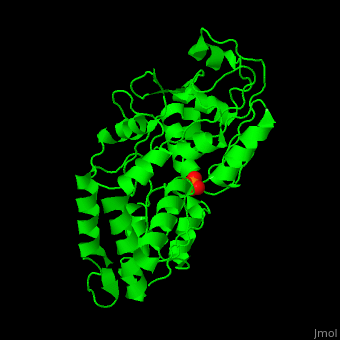
3D structures of phosphate-binding protein
Updated on 08-September-2016
1qui, 1oib - EcPBP (mutant) - Escherichia coli
4jwo – PBP – Planctomyces limnophilus
4exl, 4h1x – SpPBP – Streptococcus pneumoniae
1pbp, 2abh, 1ixh – EcPBP + Pi
1qui - EcPBP (mutant) + Br + Pi
1quj, 1qul - EcPBP (mutant) + Cl + Pi
1quk, 1ixi, 1ixg, 1a40 - EcPBP (mutant) + Pi
1a54, 1a55 - EcPBP (mutant) + dihydrogenphosphate
2v3q – hPBP + Pi – human
1pc3, 4lvq – PBP + Pi – Mycobacterium tuberculosis
3w9v, 3w9w – upPBP + Pi – unidentified prokaryote
4lat – SpPBP + Pi
4m1v - upPBP (mutant) + Pi
4omb, 4pqj – PBP + Pi – Pseudomonas aeruginosa
References
- ↑ Gonzalez D, Richez M, Bergonzi C, Chabriere E, Elias M. Crystal structure of the phosphate-binding protein (PBP-1) of an ABC-type phosphate transporter from Clostridium perfringens. Sci Rep. 2014 Oct 16;4:6636. doi: 10.1038/srep06636. PMID:25338617 doi:http://dx.doi.org/10.1038/srep06636
- ↑ Wang Z, Choudhary A, Ledvina PS, Quiocho FA. Fine tuning the specificity of the periplasmic phosphate transport receptor. Site-directed mutagenesis, ligand binding, and crystallographic studies. J Biol Chem. 1994 Oct 7;269(40):25091-4. PMID:7929197
