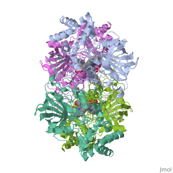
2cag
From Proteopedia
| |||||||
| , resolution 2.7Å | |||||||
|---|---|---|---|---|---|---|---|
| Sites: | and | ||||||
| Ligands: | |||||||
| Activity: | Catalase, with EC number 1.11.1.6 | ||||||
| Coordinates: | save as pdb, mmCIF, xml | ||||||
CATALASE COMPOUND II
Overview
Various enzymes use semi-stable ferryl intermediates and free radicals during their catalytic cycle, amongst them haem catalases. Structures for two transient intermediates (compounds I and II) of the NADPH-dependent catalase from Proteus mirabilis (PMC) have been determined by time-resolved X-ray crystallography and single crystal microspectrophotometry. The results show the formation and transformation of the ferryl group in the haem, and the unexpected binding of an anion during this reaction at a site distant from the haem.
About this Structure
2CAG is a Single protein structure of sequence from Proteus mirabilis. The following page contains interesting information on the relation of 2CAG with [Catalase]. Full crystallographic information is available from OCA.
Reference
Ferryl intermediates of catalase captured by time-resolved Weissenberg crystallography and UV-VIS spectroscopy., Gouet P, Jouve HM, Williams PA, Andersson I, Andreoletti P, Nussaume L, Hajdu J, Nat Struct Biol. 1996 Nov;3(11):951-6. PMID:8901874
Page seeded by OCA on Thu Mar 20 16:13:00 2008
Categories: Catalase | Proteus mirabilis | Single protein | Gouet, P. | Hajdu, J. | Jouve, H M. | HEM | Heme | Hydrogen peroxide | Iron | Nadp | Oxidoreductase (h2o2 acceptor) | Peroxidase
