Sandbox Reserved 1475
From Proteopedia
</StructureSection>
| This Sandbox is Reserved from November 5 2018 through January 1, 2019 for use in the course "CHEM 4923: Senior Project taught by Christina R. Bourne at the University of Oklahoma, Norman, USA. This reservation includes Sandbox Reserved 1471 through Sandbox Reserved 1478. |
To get started:
More help: Help:Editing |
This page is reserved for Marisha
Contents |
Retinal Dehydrogenase Type Two Structure
|
This molecular structure is Retinal Dehydrogenase Type Two (RalDH2). This enzyme is part of the super family Aldehyde Dehydrogenase. The function of this enzyme is to catalyze the oxidation of retinal to retinoic acid. Retinoic acid produces a putative morphogen that initiates pattern formation in the early embryo.[1] This is the last step in the formation of the hormone from Vitamin A (retinol).[1] Vitamin A that has been metabolized can produce retinoid derivatives which function in either vision or growth and development. [2] RalDH2 is expressed in Escherichia coli strain BL21(DE3) [3]
Function
The main function of this enzyme is to Retinoic acid. RalDH2 requires (NAD+) as a cofactor.[1] In the oxidoreductase reaction, NAD+ acts as an electron acceptor. The reaction of this enzyme is [(retinal) + (NAD+) + (H2O) ↔ (retinoic acid) + (NADH) + (H+) ]. Once the NAD+ is bound, hydrogen bonds form with non-polar residues and one basic Lysine residue. Chloride ions participate in hydrophobic interactions with Arginine residues.[1] Theres interactions cause a structural change to occur in the RalDH2 enzyme which causes it to form a more favorable folded confirmation. In the enzyme a large binding cavity is formed. tructural changes occur to stabilize the tertiary structure of RalDH2
Disease
Relevance
Structural highlights
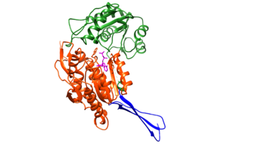
The structure was based on the mitochondrial aldehyde dehydrogenase type two. RalDH2 in a monomer made up of 3 domains: a nucleotide-binding domain (1-136, 161-270), a catalytic domain (271-484), and a tetramerization domain (137-160, 485-484) as shown in Figure 1.[1]
The tetramer can be envisioned as an "X", with nucleotide-binding sites at the tips of the "X", and the tetramerization domains as the equatorial portion of the "X".
| |||||||||||
