Main Page

From Proteopedia

Revision as of 16:10, 25 January 2019 by Joel L. Sussman (Talk | contribs)
Jump to: navigation, search
ISSN 2310-6301

As life is more than 2D, Proteopedia helps to bridge the gap between 3D structure & function of biomacromolecules

Proteopedia presents this information in a user-friendly way as a collaborative & free 3D-encyclopedia of proteins & other biomolecules.


Selected Pages In Journals Education
About this image
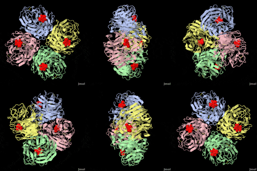
Avian Influenza Neuraminidase

Eric Martz
The first new influenza virus to emerge as an imminent pandemic threat in the 21st century is H1N1 swine flu. The drug oseltamivir (Tamiflu®) inhibits flu neuraminidase, a component necessary for virus spread, in susceptible flu strains. The development of oseltamivir was guided, in part, by crystallographically determined structures of flu neuraminidase, which is a homotetramer, shown with oseltamivir bound. Oseltamivir was designed to fit N2/N9 (neuraminidases from other strains of flu). Serendipitously, it also fits N1 by induced fit.

>>> Visit this page >>>

About this image
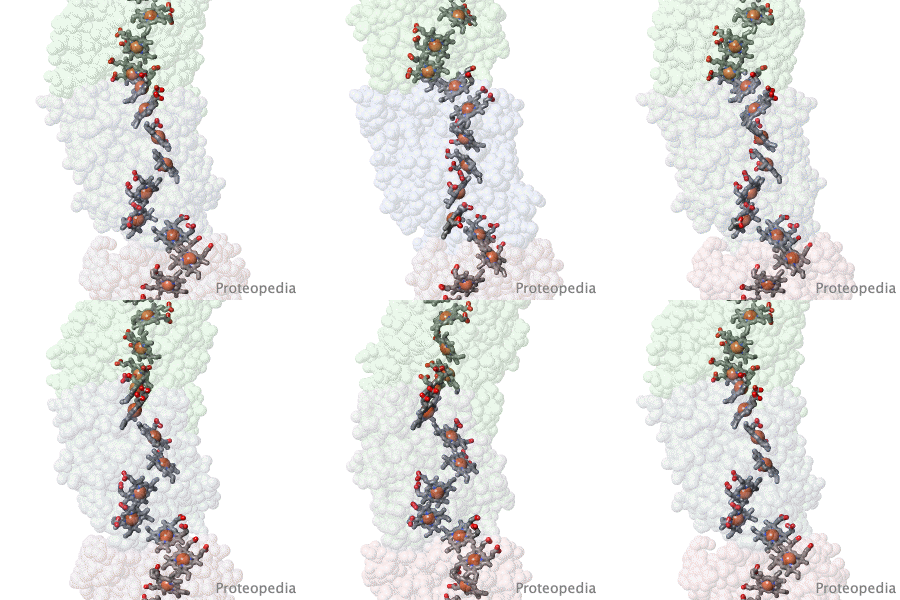
Geobacter nanowire structure surprise.

F Wang, Y Gu, JP O'Brien, SM Yi, SE Yalcin, V Srikanth, C Shen, D Vu, NL Ing, AI Hochbaum, EH Egelman, NS Malvankar. Cell 2019 doi: 10.1016/j.cell.2019.03.029
Bacteria living in anaerobic environments (no oxygen) need alternative electron acceptors in order to get energy from their food. An acceptor abundant in the earth's crust is red iron oxide ("rust"), which gets reduced to black iron oxide (magnetite). Many bacteria, such as Geobacter, get their metabolic energy by transferring electrons to acceptors that are multiple cell diameters distant, using protein nanowires. These were long thought to be pili. But when the structure of the nanowires was solved in 2019, to everyone's surprise, they turned out to be unprecedented linear polymers of multi-heme cytochromes. The hemes form an electrically conductive chain in the cores of these nanowires.

>>> Visit I3DC Interactive Visualizations >>>

About this image
Virus Capsid Geometry

The Capsid of a virus is its outer shell or "skin". Viruses have evolved intricate and elegant ways to assemble capsid protein chains into complete, usually spherical capsids, often with icosahedral symmetry. Pictured is an extremely simplified model of a capsid, where a single enlarged atom represents each of the 360 protein chains in the capsid of the Simian Virus 40 (SV40), a member of a group of cancer-causing viruses that has been extensively researched for decades.

>>> See more animations and explanation >>>

How to add content to Proteopedia

Video Guides

Who knows ...

About Interactive 3D Complements - I3DCs

List of I3DCs

How to get an I3DC for your paper

Teaching strategies using Proteopedia

Examples of pages for teaching

How to add content to Proteopedia

About Contact Hot News Table of Contents Structure Index Help

Proteopedia Page Contributors and Editors (what is this?)

Joel L. Sussman, Jaime Prilusky

Personal tools