1dkf
From Proteopedia
| |||||||
| , resolution 2.50Å | |||||||
|---|---|---|---|---|---|---|---|
| Ligands: | , | ||||||
| Related: | 1LBD, 2LBD
| ||||||
| Resources: | FirstGlance, OCA, PDBsum, RCSB | ||||||
| Coordinates: | save as pdb, mmCIF, xml | ||||||
CRYSTAL STRUCTURE OF A HETERODIMERIC COMPLEX OF RAR AND RXR LIGAND-BINDING DOMAINS
Overview
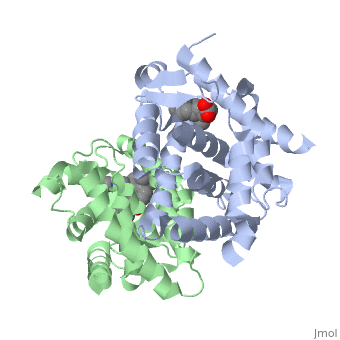
The crystal structure of a heterodimer between the ligand-binding domains (LBDs) of the human RARalpha bound to a selective antagonist and the constitutively active mouse RXRalphaF318A mutant shows that, pushed by a bulky extension of the ligand, RARalpha helix H12 adopts an antagonist position. The unexpected presence of a fatty acid in the ligand-binding pocket of RXRalpha(F318A is likely to account for its apparent "constitutivity." Specific conformational changes suggest the structural basis of pure and partial antagonism. The RAR-RXR heterodimer interface is similar to that observed in most nuclear receptor (NR) homodimers. A correlative analysis of 3D structures and sequences provides a novel view on dimerization among members of the nuclear receptor superfamily.
About this Structure
1DKF is a Protein complex structure of sequences from Homo sapiens and Mus musculus. Full crystallographic information is available from OCA.
Reference
Crystal structure of a heterodimeric complex of RAR and RXR ligand-binding domains., Bourguet W, Vivat V, Wurtz JM, Chambon P, Gronemeyer H, Moras D, Mol Cell. 2000 Feb;5(2):289-98. PMID:10882070
Page seeded by OCA on Sun Mar 30 19:43:16 2008
Categories: Homo sapiens | Mus musculus | Protein complex | Bourguet, W. | Chambon, P. | Gronemeyer, H. | Moras, D. | SPINE, Structural Proteomics in Europe. | Vivat, V. | Wurtz, J M. | Helical sandwich | Heterodimer | Hormone/growth factor receptor | Protein-ligand complex | Spine | Structural genomic | Structural proteomics in europe
