The CFTR is a chloride channel, and is regulated by PKA phosphorylation, cAMP levels, and ATP/ADP ratios. Mutations in the CFTR cause the disease cystic fibrosis.
One feature of the CFTR is a Walker motif, which is found in ATP binding proteins.It is also known as a P (or phosphate binding) loop.
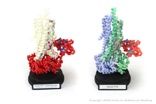
3D Printed Physical Model of the CFTR protein
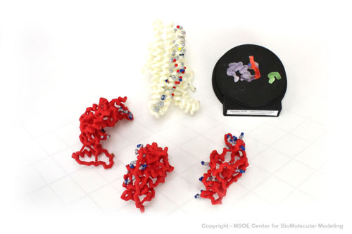
Shown below are 3D printed physical models of the Cystic Fibrosis Transmembrane Conductance Regulator (CFTR) protein. The backbone model on the left is colored by region, with the transmembrane domain white and the atp-binding and regulatory domains colored red. The backbone model on the right is colored by regional repeat, with the first repeat blue, the second repeat green and the regulatory domain colored red. The models have been designed with embedded magnets to disassemble into the key regions of the structure.
The MSOE Center for BioMolecular Modeling
The MSOE Center for BioMolecular Modeling uses 3D printing technology to create physical models of protein and molecular structures, making the invisible molecular world more tangible and comprehensible. To view more protein structure models, visit our Model Gallery.


