Uridine phosphorylase
From Proteopedia
| |||||||||||
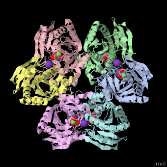
3D Structures of uridine phosphorylase
Updated on 23-March-2020
References
- ↑ Pizzorno G, Cao D, Leffert JJ, Russell RL, Zhang D, Handschumacher RE. Homeostatic control of uridine and the role of uridine phosphorylase: a biological and clinical update. Biochim Biophys Acta. 2002 Jul 18;1587(2-3):133-44. PMID:12084455
- ↑ Renck D, Santos AA Jr, Machado P, Petersen GO, Lopes TG, Santos DS, Campos MM, Basso LA. Human uridine phosphorylase-1 inhibitors: a new approach to ameliorate 5-fluorouracil-induced intestinal mucositis. Invest New Drugs. 2014 Dec;32(6):1301-7. doi: 10.1007/s10637-014-0135-0. Epub, 2014 Jul 23. PMID:25052233 doi:http://dx.doi.org/10.1007/s10637-014-0135-0
- ↑ Lashkov AA, Sotnichenko SE, Prokofiev II, Gabdulkhakov AG, Agapov II, Shtil AA, Betzel C, Mironov AS, Mikhailov AM. X-ray structure of Salmonella typhimurium uridine phosphorylase complexed with 5-fluorouracil and molecular modelling of the complex of 5-fluorouracil with uridine phosphorylase from Vibrio cholerae. Acta Crystallogr D Biol Crystallogr. 2012 Aug;68(Pt 8):968-74. Epub 2012 Jul 17. PMID:22868762 doi:http://dx.doi.org/10.1107/S090744491201815X
