Overview
The PWWP domain belongs to the Royal family, which consists of Tudor, chromodomain, MBT(Malignant Brain Tumor), and PWWP domains. The PWWP domain was first identified as a structural motif includes 100 to 130 amino acids in WHSC1 protein research. The researchers named after its very conservative sequence, Pro-Trp-Trp-Pro located at the β2 strand of protein.[1] This domain is mainly related to the recognition of methylated histone tail lysine and related to the epigenetic regulation of genes. BRPF1 PWWP domain, which has very similar structure with 3pfs, presents on actively transcribing cells and may regulate Hox A9 gene which is related to MOZ-TIF2-dependant leukemia.[2] The 3pfs motif is relatively mystery we may assume that 3pfs also do similar works
Structure
Two disticntive The first proline affects the stability and aggregation of the protein. The proline gives more stability and oligomerization to the protein, compared to the alanine at the same position.[3] It has two distinctive substructural motifs: β-barrel at the N-terminal region and helixes at C-terminal region.[4] The β-barrel is a very conservative feature of PWWP domains and it is composed of 5 β-strands. 3pfs has 3 helixes which are little more than many of its relatives. The stability of domain comes from both inter-substructure and intra-substructure interactions including hydrogen bonds and polar interactions.
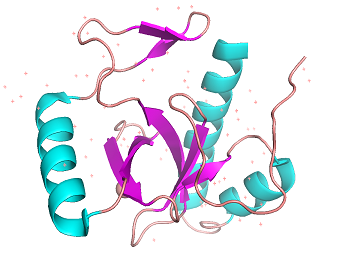
Figure 1: Cartoon model of 3PFS motif. β-sheets(magenta) and helixes(orange) are shown. generated in PyMOL using PDB:
3PFS Function
Nonspecific binding
The PWWP domains have a significant amount of basic residues.[5] 3pfs also has 11 lysine residues and 12 arginine residues. These residues' sidechains would be positively charged on in vivo pH. The DNA strand phosphate backbone, which is negatively charged, is attracted to PWWP domain surface due to the nature of attraction between the opposite charges.
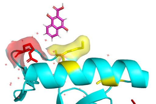
Figure 2: Lysine(yellow) and Arginine(red) residues on 3PFS surface interacting with DNA backbone generated in PyMOL using PDB:
3PFS Methyl-lysine reader
PWWP domains are one of epigenetic regulators which recognize specific lysine residues which are methylated. Even though there is no detailed research on 3pfs, it is assumed to have similar function with BRPF1 PWWP domain which is shown to have methylated histone binding activity.[6] With a high-throughput mass spectrometry screening, this motif is suggested as an essential motif of histone 3 lysine 36 trimethylation binding.[7] The β-strands and Pro-Trp-Trp-Pro motif form hydrophobic cavity, which is the binding site of methylated lysines.[8] Since the aromatic cage is a common structure in the Royal family, it can be a common tool for methylated lysine binding.[9]
Evolutionary conservations
PWWP domain is only found in eukaryotes, from a yeast to a human. The number of PWWP containing protein is also varying on species; the human has 20. The loyal family has a common structural characteristic, three conserved β-strands.[10] The PWWP domain is also believed to be evolved from a common ancestor which had 3 β-strands. The most conservative section is the PWWP sequence, which it is named after.
Therapheutic features
There is no research on the relationship between BRPF3 PWWP domain and any human disease. However, its sibling gene BRPF2 PWWP domain is associated with schizophrenia and bipolar affective disorder.[11]
This is a sample scene created with SAT to by Group, and another to make of the protein. You can make your own scenes on SAT starting from scratch or loading and editing one of these sample scenes.


