Function
Pyruvate dehydrogenase kinase (PDK) is part of the pyruvate dehydrogenase complex. This complex is located in the mitochondria and converts pyruvate to acetyl-CoA as part of the citric acid cycle. PDK phosphphorylates serine residues on pyruvate dehydrogenase using ATP. There are 4 isozymes of PDK. The isozymes differ in length, activity and phosphorylation sites[1]. PDK1 is abundant in heart cells. PDK2 is abundant in mitochondria. PDK3 is abundant in testis. PDK4 is abundant in muscle and heart.
Relevance
Inhibition of PDK decreases the damage caused by heart ischemia and are used in diabetes and cancer patients[2][3].
Structural highlights
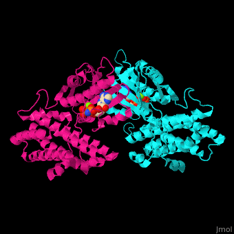
[4]. Water molecules are shown as red spheres. .
3D structures of pyruvate dehydrogenase kinase
Updated on 14-October-2020
References
- ↑ Korotchkina LG, Patel MS. Site specificity of four pyruvate dehydrogenase kinase isoenzymes toward the three phosphorylation sites of human pyruvate dehydrogenase. J Biol Chem. 2001 Oct 5;276(40):37223-9. Epub 2001 Aug 2. PMID:11486000 doi:10.1074/jbc.M103069200
- ↑ Roche TE, Hiromasa Y. Pyruvate dehydrogenase kinase regulatory mechanisms and inhibition in treating diabetes, heart ischemia, and cancer. Cell Mol Life Sci. 2007 Apr;64(7-8):830-49. PMID:17310282 doi:10.1007/s00018-007-6380-z
- ↑ Sutendra G, Michelakis ED. Pyruvate dehydrogenase kinase as a novel therapeutic target in oncology. Front Oncol. 2013 Mar 7;3:38. doi: 10.3389/fonc.2013.00038. eCollection 2013. PMID:23471124 doi:http://dx.doi.org/10.3389/fonc.2013.00038
- ↑ Kukimoto-Niino M, Tokmakov A, Terada T, Ohbayashi N, Fujimoto T, Gomi S, Shiromizu I, Kawamoto M, Matsusue T, Shirouzu M, Yokoyama S. Inhibitor-bound structures of human pyruvate dehydrogenase kinase 4. Acta Crystallogr D Biol Crystallogr. 2011 Sep;67(Pt 9):763-73. doi:, 10.1107/S090744491102405X. Epub 2011 Aug 9. PMID:21904029 doi:10.1107/S090744491102405X