This is a default text for your page Natalia de Araujo./Sandbox 1. Click above on edit this page to modify. Be careful with the < and > signs.
You may include any references to papers as in: the use of JSmol in Proteopedia [1] or to the article describing Jmol [2] to the rescue.
The first structure loaded on the left-hand side of the screen corresponds to 4jon, the Crystal structure of CEP170's transcript variant beta, from Homo sapiens. 4jon is a 5 chain structure obtained by X-ray Crystallography [1] and represents the first 126 residues of CEP170's N-termini region.
The second structure available for CEP170 is , a predicted 3D model.
Here we are going to focus on the Q5SW79 model due to its full sequence, allowing us to highlight residues and regions important for the understanding of CEP170's functions and interactions.
|
|
| Available 3D models: | 4jon, |
| Specie: | Human |
| Gene: | CEP170, FAM68A, KAB, KIAA0470, NM_001042404 (HUMAN) |
| Splice Variants | CEP170α, CEP170β, and CEP170γ |
| RefSeq (mRNA): | CEP170α: NM_014812[2], CEP170β: NM_001042404 [3], CEP170γ: NM_001042405[4] |
| RefSeq (protein): | CEP170α: NP_055627[5], CEP170β: NP_001035863[6], CEP170γ: NP_001035864[7] |
| UniProt: | Q5SW79[8] |
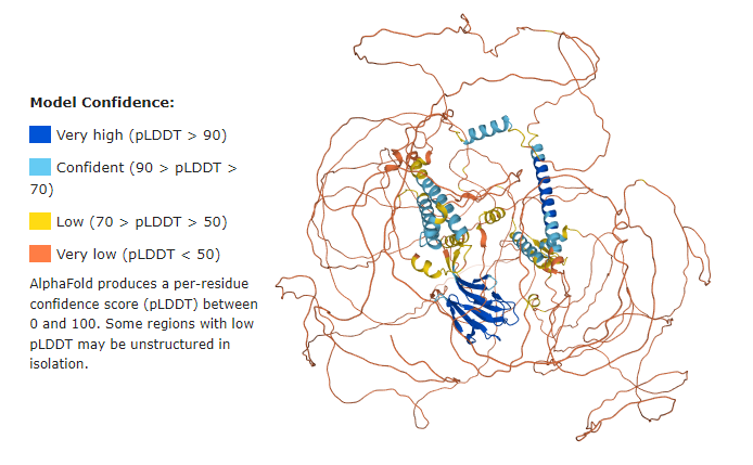
About the Q5SW79 model
As seen below, the majority of the Q5SW79 structure has very low confidence, meaning the model most probably does not represent the real protein structure.
However, it stills helps us to have a good idea of the whole protein.
CEP170 gene
The CEP170 gene (OMIM#613023[9]) spans over XX kb at 1q43, contains XX exons, and encodes the Centrosomal Protein of 170 kDa (CEP170).
Function
Microtubule organization
Microtubule nucleation
Protein-Protein Interactions
- CHEK1 and circCHEK1_246aa
Through Co-IP [10]and MS[11] CEP170 was identified as a putative interactor with CHEK1, further confirmed with Co-IP in cells overexpressing CHEK1 [3]
CHEK and circCHEK1_246aa (isoform from a cRNA) phosphorylate the residue of CEP170, which results in chromosomal instability seen in multiple myeloid cells.
Structural highlights
As seen by Guarguaglini et al. (2005) all three splice variants harbor an N-terminal forkhead-associated (FHA) domain and a serine-rich domain and a short coiled-coil region in the C-terminal half.
The C-terminus region harbors 3 globular domains, where one is located at and the second at
Disease
This is a sample scene created with SAT to by Group, and another to make of the protein. You can make your own scenes on SAT starting from scratch or loading and editing one of these sample scenes.