This is a default text for your page Camryn Jiles/Sandbox 1. Click above on edit this page to modify. Be careful with the < and > signs.
You may include any references to papers as in: the use of JSmol in Proteopedia [1] or to the article describing Jmol [2] to the rescue.
Introduction
Orexin, also known as hypocretin, are a group of neuropeptides identified in the late 1990’s, located in the lateral region of the hypothalamus. Neuropeptides are short strings of amino acids that are synthesized and released by neurons in the brain. These chemical messengers will typically bind to G protein-coupled receptors (GPCRs) to regulate neural activity. Human neuropeptide Y consists of three ligands (NPY, PYY and PP) and four receptors (Y1R, Y2R, Y3R, Y4R and Y5R). NPY is abundant in the brain and its structure consists of a C-terminal segment that forms an α-helix; its N terminal is flexible and relatively unstructured. The receptors act as an antagonist, when the ligand and receptor come together forming a complex, resulting in an extracellular conformational change. The C terminus of NPY will penetrate the transmembrane core of the receptor which in turn, activates the G protein-coupled receptor. One key characteristic that is indicative of this type of activated GPCR includes interaction “between R138 of the (D/E)R(Y/H) motif and Y231 and Y320 of the NPY motif” (Park 2).
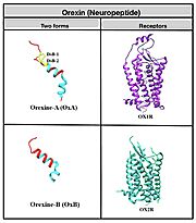
Orexin's primarily play a role in the central nervous system and are responsible for arousal, wakefulness and appetite. Additionally, they are capable of various physiology functions, regulating reproductive and neuroendocrine functions, gastrointestinal motility, blood pressure, metabolism and energy balance. Orexin consists of 131 amino acids and is broken down into two forms: orexin-A (OxA) which consists of 33 amino acids and orexin-B (OxB), consisting of 28 amino acids. OxA consists of a pyroglutamyl residue on the N terminus, two disulfide bridges and an amidated C-terminus. OxB also contains an amidated C-terminus. Both proteins also contain two ɑ-helices within their structure. OxA and OxB activates orexin receptor type 1(OX1R) and orexin receptor type 2 (OXR2) that act on G protein-coupled receptors located on effector cells. It is worth noting that OX1R has a better affinity for OxA and OX2R for OxB. This signaling pathway and resulting influx of calcium into the intracellular space, is what allows orexin to mediate the various biological actions across the central and peripheral nervous system.
Function & Structure
Relevance
Inhibition
Conclusion
This is a sample scene created with SAT to by Group, and another to make of the protein. You can make your own scenes on SAT starting from scratch or loading and editing one of these sample scenes.


