We apologize for Proteopedia being slow to respond. For the past two years, a new implementation of Proteopedia has been being built. Soon, it will replace this 18-year old system. All existing content will be moved to the new system at a date that will be announced here.
ExbD
From Proteopedia
| |||||||||||
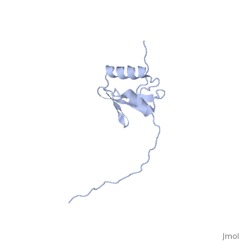

3D structures of ExbD
2pfu – EcExbD periplasmic domain – Escherichia coli – NMR
5sv1 – EcExbD residues 1-49 + EcExbB
6tyi – EcExbD + ExbB – Cryo EM
References
- ↑ 1.0 1.1 1.2 Kampfenkel K, Braun V. Membrane topology of the Escherichia coli ExbD protein. J Bacteriol. 1992 Aug;174(16):5485-7. PMID:1644779
- ↑ Held KG, Postle K. ExbB and ExbD do not function independently in TonB-dependent energy transduction. J Bacteriol. 2002 Sep;184(18):5170-3. PMID:12193634
- ↑ Braun V, Herrmann C. Point mutations in transmembrane helices 2 and 3 of ExbB and TolQ affect their activities in Escherichia coli K-12. J Bacteriol. 2004 Jul;186(13):4402-6. PMID:15205446 doi:10.1128/JB.186.13.4402-4406.2004
