Structure
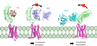
There are 3 main domains of the Thyroid Stimulating Hormone Receptor. First is the shown in green. Region is concave in shape and is made up of primarily beta sheets and rich in Leucines. It is also called the leucine rich region. This is also the domain for key Lysine residues, which play a key role in binding. Second is the shown in pink. This domain is made up of 7 helices and undergoes a conformation change upon ligand binding that activates the GPCR signal cascade (GPCR is shown in yellow). The Third region of the TSHR is the shown in orange. The Hinge Region plays a key role in the movement and stability of the TSHR.
Binding of Thyroid Stimulating Hormone to TSHR
by complementary shape and several polar/nonpolar interactions
Words...
words...
words....
Words....
Blocking TSHR in Active/Inactive States
Structural highlights
This is a sample scene created with SAT to by Group, and another to make of the protein. You can make your own scenes on SAT starting from scratch or loading and editing one of these sample scenes.

