Protein disulfide oxidoreductase
From Proteopedia
| |||||||||||
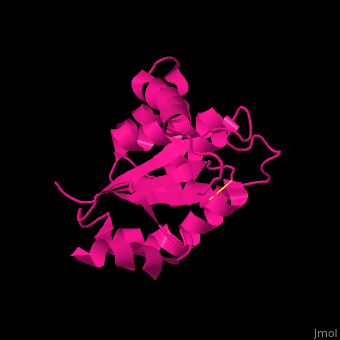
3D structures of protein disulfide oxidoreductase
Updated on 08-October-2020
1a8l – PDOR – Pyrococcus furiosus
1bq7 – EcDsbA (mutant) – Escherichia coli
2hi7, 3e9j, 2zup - EcDsbA (mutant) + EcDsbB (mutant)
2leg - EcDsbA (mutant) + EcDsbB (mutant) - NMR
1fo5 – PDOR – Methanocaldococcus jannaschii – NMR
2ayt – PDOR – Aquifex aeolicus
2hls – PDOR – Aeropyrus pernix
2rem - DsbA + peptide – Xylella fastidiosa
1st9, 1su9, 2f9s, 2h1d, 3gha – BsResA soluble domain – Bacillus subtilis
3eu3 – BsBdbB
2h19, 2h1a, 2h1b, 2h1g, 3c71, 3c73 - BsResA soluble domain (mutant)
4k6x – PDOR – Mycobacterium tuberculosis
3bci – SaDsbA – Staphylococcus aureus
3bck, 3bd2 - SaDsbA (mutant)
3gh9 - SaBdbB
3erw – SaPDOR sporulation
3fkf – PDOR – Bacterioides fragilis
1bed – DsbA – Vibrio cholerae
4z7x - PDOR - Actinomyces oris
5cnw, 5e59 - DrPDOR - Deinicoccus radiodurans
5cp1 - DrPDOR (mutant)
4mcu - PDOR - Klebsiella pneumoniae
4oce - PDOR - Proteus mirabillis
4jrr - DsbA1 - Legionella pneumophila
For DsbB see Thiol:disulfide interchange protein.
References
- ↑ Pedone E, Ren B, Ladenstein R, Rossi M, Bartolucci S. Functional properties of the protein disulfide oxidoreductase from the archaeon Pyrococcus furiosus: a member of a novel protein family related to protein disulfide-isomerase. Eur J Biochem. 2004 Aug;271(16):3437-48. PMID:15291821 doi:http://dx.doi.org/10.1111/j.0014-2956.2004.04282.x
- ↑ Heras B, Kurz M, Jarrott R, Byriel KA, Jones A, Thony-Meyer L, Martin JL. Expression and crystallization of DsbA from Staphylococcus aureus. Acta Crystallogr Sect F Struct Biol Cryst Commun. 2007 Nov 1;63(Pt, 11):953-6. Epub 2007 Oct 24. PMID:18007049 doi:10.1107/S174430910704821X
