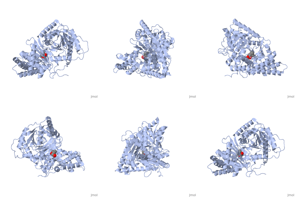
1s5o
From Proteopedia
| |||||||
| , resolution 1.8Å | |||||||
|---|---|---|---|---|---|---|---|
| Ligands: | |||||||
| Activity: | Carnitine O-acetyltransferase, with EC number 2.3.1.7 | ||||||
| Resources: | FirstGlance, OCA, PDBsum, RCSB | ||||||
| Coordinates: | save as pdb, mmCIF, xml | ||||||
Structural and Mutational Characterization of L-carnitine Binding to Human carnitine Acetyltransferase
Overview
We report the crystal structure of a binary complex of human peroxisomal carnitine acetyltransferase and the substrate l-carnitine, refined to a resolution of 1.8 Angstrom with an R(factor) value of 18.9% (R(free)=22.3%). L-carnitine binds to a preformed pocket in the active site tunnel of carnitine acetyltransferase aligned with His(322). The quaternary nitrogen of carnitine forms a pi-cation interaction with Phe(545), while Arg(497) forms an electrostatic interaction with the negatively charged carboxylate group. An extensive hydrogen bond network also occurs between the carboxylate group and Tyr(431), Thr(444), and a bound water molecule. Site-directed mutagenesis and kinetic characterization reveals that Tyr(431), Thr(444), Arg(497), and Phe(545) are essential for high affinity binding of L-carnitine.
About this Structure
1S5O is a Single protein structure of sequence from Homo sapiens. Full crystallographic information is available from OCA.
Reference
Structural and mutational characterization of L-carnitine binding to human carnitine acetyltransferase., Govindasamy L, Kukar T, Lian W, Pedersen B, Gu Y, Agbandje-McKenna M, Jin S, McKenna R, Wu D, J Struct Biol. 2004 Jun;146(3):416-24. PMID:15099582
Page seeded by OCA on Sun Mar 30 23:37:44 2008
Categories: Carnitine O-acetyltransferase | Homo sapiens | Single protein | Agbandje-Mckenna, M. | Govindasamy, L. | Gu, Y. | Jin, S. | Kukar, T. | Lian, W. | Mckenna, R. | Pedersen, B. | Wu, D. | Binary complex | Carnitine acetyltransferase | Steady-state enzyme kinetic | Substrate binding site | X-ray structure

