Ephrin
From Proteopedia
| |||||||||||
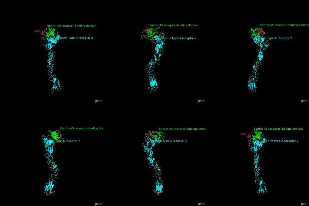
3D structures of ephrin
Updated on 18-June-2024
References
- ↑ Egea J, Klein R. Bidirectional Eph-ephrin signaling during axon guidance. Trends Cell Biol. 2007 May;17(5):230-8. Epub 2007 Apr 8. PMID:17420126 doi:http://dx.doi.org/10.1016/j.tcb.2007.03.004
- ↑ Hoshino N, Altarshan Y, Alzein A, Fernando AM, Nguyen HT, Majewski EF, Chen VC, Rochlin MW, Yu WM. Ephrin-A3 is required for tonotopic map precision and auditory functions in the mouse auditory brainstem. J Comp Neurol. 2021 Nov;529(16):3633-3654. PMID:34235739 doi:10.1002/cne.25213
- ↑ Frisén J, Yates PA, McLaughlin T, Friedman GC, O'Leary DD, Barbacid M. Ephrin-A5 (AL-1/RAGS) is essential for proper retinal axon guidance and topographic mapping in the mammalian visual system. Neuron. 1998 Feb;20(2):235-43. PMID:9491985 doi:10.1016/s0896-6273(00)80452-3
- ↑ Himanen JP, Yermekbayeva L, Janes PW, Walker JR, Xu K, Atapattu L, Rajashankar KR, Mensinga A, Lackmann M, Nikolov DB, Dhe-Paganon S. Architecture of Eph receptor clusters. Proc Natl Acad Sci U S A. 2010 May 26. PMID:20505120

