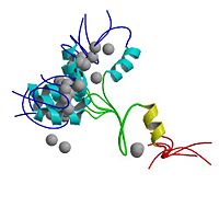
2adr
From Proteopedia
| |||||||
| Sites: | |||||||
| Ligands: | |||||||
| Resources: | FirstGlance, OCA, PDBsum, RCSB | ||||||
| Coordinates: | save as pdb, mmCIF, xml | ||||||
ADR1 DNA-BINDING DOMAIN FROM SACCHAROMYCES CEREVISIAE, NMR, 25 STRUCTURES
Overview
The region responsible for sequence-specific DNA binding by the transcription factor ADR1 contains two Cys2-His2 zinc fingers and an additional N-terminal proximal accessory region (PAR). The N-terminal (non-finger) PAR is unstructured in the absence of DNA and undergoes a folding transition on binding the DNA transcription target site. We have used a set of HN-HN NOEs derived from a perdeuterated protein-DNA complex to describe the fold of ADR1 bound to the UAS1 binding site. The PAR forms a compact domain consisting of three antiparallel strands that contact A-T base pairs in the major groove. The three-strand domain is a novel fold among all known DNA-binding proteins. The PAR shares sequence homology with the N-terminal regions of other zinc finger proteins, suggesting that it represents a new DNA-binding module that extends the binding repertoire of zinc finger proteins.
About this Structure
2ADR is a Single protein structure of sequence from Saccharomyces cerevisiae. Full crystallographic information is available from OCA.
Reference
A folding transition and novel zinc finger accessory domain in the transcription factor ADR1., Bowers PM, Schaufler LE, Klevit RE, Nat Struct Biol. 1999 May;6(5):478-85. PMID:10331877
Page seeded by OCA on Mon Mar 31 01:51:17 2008

