1bm0
From Proteopedia
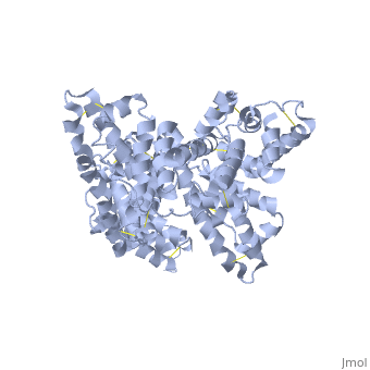

CRYSTAL STRUCTURE OF HUMAN SERUM ALBUMIN
Overview
A new triclinic crystal form of human serum albumin (HSA), derived either from pool plasma (pHSA) or from a Pichia pastoris expression system (rHSA), was obtained from polyethylene glycol 4000 solution. Three-dimensional structures of pHSA and rHSA were determined at 2.5 A resolution from the new triclinic crystal form by molecular replacement, using atomic coordinates derived from a multiple isomorphous replacement work with a known tetragonal crystal form. The structures of pHSA and rHSA are virtually identical, with an r.m. s. deviation of 0.24 A for all Calpha atoms. The two HSA molecules involved in the asymmetric unit are related by a strict local twofold symmetry such that the Calpha atoms of the two molecules can be superimposed with an r.m.s. deviation of 0.28 A in pHSA. Cys34 is the only cysteine with a free sulfhydryl group which does not participate in a disulfide linkage with any external ligand. Domains II and III both have a pocket formed mostly of hydrophobic and positively charged residues and in which a very wide range of compounds may be accommodated. Three tentative binding sites for long-chain fatty acids, each with different surroundings, are located at the surface of each domain.
About this Structure
1BM0 is a Single protein structure of sequence from Homo sapiens. Full crystallographic information is available from OCA.
Reference
Crystal structure of human serum albumin at 2.5 A resolution., Sugio S, Kashima A, Mochizuki S, Noda M, Kobayashi K, Protein Eng. 1999 Jun;12(6):439-46. PMID:10388840 Page seeded by OCA on Fri May 2 11:41:00 2008
