7rsa
From Proteopedia
| |||||||||
| 7rsa, resolution 1.26Å () | |||||||||
|---|---|---|---|---|---|---|---|---|---|
| Ligands: | , | ||||||||
| Activity: | Pancreatic ribonuclease, with EC number 3.1.27.5 | ||||||||
| |||||||||
| |||||||||
| |||||||||
| Resources: | FirstGlance, OCA, RCSB, PDBsum | ||||||||
| Coordinates: | save as pdb, mmCIF, xml | ||||||||
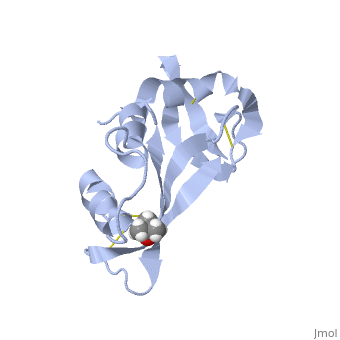

STRUCTURE OF PHOSPHATE-FREE RIBONUCLEASE A REFINED AT 1.26 ANGSTROMS
Overview
The structure of phosphate-free bovine ribonuclease A has been refined at 1.26-A resolution by a restrained least-squares procedure to a final R factor of 0.15. X-ray diffraction data were collected with an electronic position-sensitive detector. The final model consists of all atoms in the polypeptide chain including hydrogens, 188 water sites with full or partial occupancy, and a single molecule of 2-methyl-2-propanol. Thirteen side chains were modeled with two alternate conformations. Major changes to the active site include the addition of two waters in the phosphate-binding pocket, disordering of Gln-11, and tilting of the imidazole ring of His-119. The structure of the protein and of the associated solvent was extensively compared with three other high-resolution, refined structures of this enzyme.
About this Structure
7RSA is a Single protein structure of sequence from Bos taurus. Full crystallographic information is available from OCA.
Reference
Structure of phosphate-free ribonuclease A refined at 1.26 A., Wlodawer A, Svensson LA, Sjolin L, Gilliland GL, Biochemistry. 1988 Apr 19;27(8):2705-17. PMID:3401445 Page seeded by OCA on Sun May 4 22:47:35 2008
