Sandbox3
From Proteopedia
This sandbox is in use until June 1, 2009 for UMass Chemistry 490a. Others please do not edit this page. Thanks!
L Thompson 3/1/09 Start by finding a protein with a known structure that interests you (see assignment sheet instructions). Determine whether anyone else in the class has already started a sandbox page for your protein (not sure yet how to do this easily...). If so, join their page. If not, start a new sandbox page.
Copy the message at the top into your sandbox page to "reserve" this sandbox for this course (others can still edit it but there is a way to go back -- not sure how yet).
Give "credit" for all your entries: add your name (like mine above) and also the source of anything that you got from another source.
Look of the pdb id for your protein in the Protein Data Bank. Use this code to find the Proteopedia entry: this is the "automatically seeded entry". Copy this into your sandbox as a starting point.
I looked for the earliest GFP structure I could find in the protein data bank and found 1ema. I searched Proteopedia for 1ema, clicked "edit page" button at the top, and then copied the contents below.

| |||||||||
| 1ema, resolution 1.90Å () | |||||||||
|---|---|---|---|---|---|---|---|---|---|
| Non-Standard Residues: | , | ||||||||
| |||||||||
| |||||||||
| Resources: | FirstGlance, OCA, RCSB, PDBsum | ||||||||
| Coordinates: | save as pdb, mmCIF, xml | ||||||||


Contents |
GREEN FLUORESCENT PROTEIN FROM AEQUOREA VICTORIA
Template:ABSTRACT PUBMED 8703075
About this Structure
1EMA is a 1 chain structure of sequence from Aequorea victoria. The June 2003 RCSB PDB Molecule of the Month feature on Green Fluorescent Protein (GFP) by David S. Goodsell is 10.2210/rcsb_pdb/mom_2003_6. Full crystallographic information is available from OCA.
Reference
- Ormo M, Cubitt AB, Kallio K, Gross LA, Tsien RY, Remington SJ. Crystal structure of the Aequorea victoria green fluorescent protein. Science. 1996 Sep 6;273(5280):1392-5. PMID:8703075
Page seeded by OCA on Tue Feb 17 16:53:56 2009
This is a placeholder
This is a placeholder text to help you get started in placing a Jmol applet on your page. At any time, click "Show Preview" at the bottom of this page to see how it goes.
Replace the PDB id (use lowercase!) after the STRUCTURE_ and after PDB= to load and display another structure.
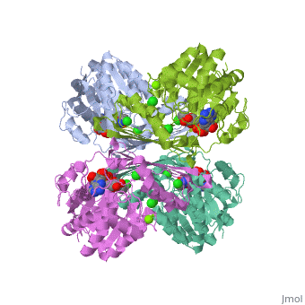
| |||||||||
| 3cin, resolution 1.70Å () | |||||||||
|---|---|---|---|---|---|---|---|---|---|
| Ligands: | , , | ||||||||
| Gene: | TM1419, TM_1419 (Thermotoga maritima MSB8) | ||||||||
| Activity: | Inositol-3-phosphate synthase, with EC number 5.5.1.4 | ||||||||
| |||||||||
| |||||||||
| Resources: | FirstGlance, OCA, RCSB, PDBsum, TOPSAN | ||||||||
| Coordinates: | save as pdb, mmCIF, xml | ||||||||