User:Eleanor Crabb/Sandbox 1
From Proteopedia
|
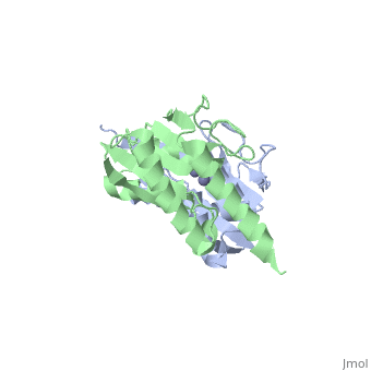
The interactive image on the right shows the protein with the pdb code 1z3a. You will note that there are two chains shown in green and blue.
In order to look at the structure of proteins in more detail we will concentrate on just a . Click on the link in green.
This is a cartoon representation of the protein. This shows the back bone of the protein. You may be more familiar with the following representations:
Let us now look at just a single length of the chain.
If we now draw a line through the backbone of the chain Let us return to the whole chain. The arrangement of the residues is the primary structure - coloured according to . Secondary structure
