File list
From Proteopedia
| Name | User | Size | Description | |
|---|---|---|---|---|
| 10:01, 2 November 2008 | ZGB4.pdb (file) | Alexander Berchansky | 682 KB | |
| 09:50, 2 November 2008 | ZGB3.pdb (file) | Alexander Berchansky | 2.05 MB | |
| 09:47, 2 November 2008 | 1zgb.3.pdb (file) | Alexander Berchansky | 2.05 MB | |
| 08:57, 2 November 2008 | 1zgb.pdb (file) | Alexander Berchansky | 580 KB | |
| 06:48, 30 October 2008 | Transparent.gif (file) | Jaime Prilusky | 807 B | |
| 14:42, 29 October 2008 | ZGBm3.pdb (file) | Alexander Berchansky | 1.08 MB | |
| 14:18, 29 October 2008 | ZGBm2.pdb (file) | Alexander Berchansky | 1.08 MB | (1zgb+1h22+1h23) |
| 11:25, 29 October 2008 | ZGBm1.pdb (file) | Alexander Berchansky | 716 KB | (1ZGB + 1ACJ) |
| 12:10, 28 October 2008 | ZGBm.pdb (file) | Alexander Berchansky | 397 KB | (1zgb modified) |
| 11:33, 28 October 2008 | A1e.pdb (file) | Alexander Berchansky | 3 KB | ((RS)-(±)-N-9-(1,2,3,4-tetrahydroacridinyl)-N'-5-[5,6,7,8-tetrahydro-2'(1'H)-quinolinonyl]-1,10-diaminodecane [(RS)-(±)-3]) |
| 05:50, 21 October 2008 | Unremediated1rpuWithPolyview3D.png (file) | Wayne Decatur | 345 KB | (Unremediated 1rpu fancy image made with Polyview-3D Settings used:</h3> Data source: <b>Custom file</b><br> Structure source: <b>OTHER</b> server<br> Show ligands<br> Ligands are rendered as <b>spheres</b><br> Initial rotation around <b>X</b> axis: <b>115) |
| 02:08, 21 October 2008 | Lacrep_anim_large.gif (file) | Eric Martz | 558 KB | (Animation of lac repressor binding to DNA, converting from non-specific binding 1osl to specific binding 1l1m, as explained at Lac repressor.) |
| 09:58, 20 October 2008 | 3d3m.png (file) | OCA | 291 KB | (Source Jena Library http://www.fli-leibniz.de/IMAGE.html) |
| 16:46, 17 October 2008 | 1osl_14_1l1m_9_morph.pdb (file) | Eric Martz | 451 KB | (Morph of 1osl model 14 (with modified DNA) to 1l1m model 9. For methods, please see Lac repressor morph methods. This is a duplicate of the file Image:1osl_19_1l1m_9_morph.pdb because I had problems loading that file.) |
| 22:21, 15 October 2008 | 1l1m_ca.pdb (file) | Eric Martz | 45 KB | (1l1m with the header, except for the HELIX records, and all ATOM records deleted, except for alpha carbon and phosphorus ATOM records.) |
| 10:42, 15 October 2008 | 2zet.png (file) | OCA | 325 KB | (Source Jena Library http://www.fli-leibniz.de/IMAGE.html) |
| 10:40, 15 October 2008 | 3dmq.png (file) | OCA | 464 KB | (Source Jena Library http://www.fli-leibniz.de/IMAGE.html) |
| 02:00, 15 October 2008 | 1osl_ca.pdb (file) | Eric Martz | 43 KB | (1osl.pdb (20 models) with all ATOM records deleted, except for alpha carbons. Header deleted, except that secondary structure (HELIX) records were retained. Gzipped. This reduced the uncompressed file from about 5 megabytes to about a quarter megabyte (~2) |
| 15:52, 8 October 2008 | Myoglobin1958.png (file) | Eric Martz | 32 KB | (Portion of Fig. 2 from Kendrew et al., Nature 181:662, 1958, with permission for use in proteopedia.org: License number 2044270781604 obtained Oct 8, 2008 by Eric Martz. Credit line to read: Adapted by permission from Macmillan Publishers Ltd: Nature 181:) |
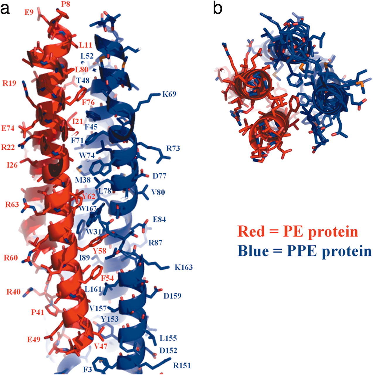
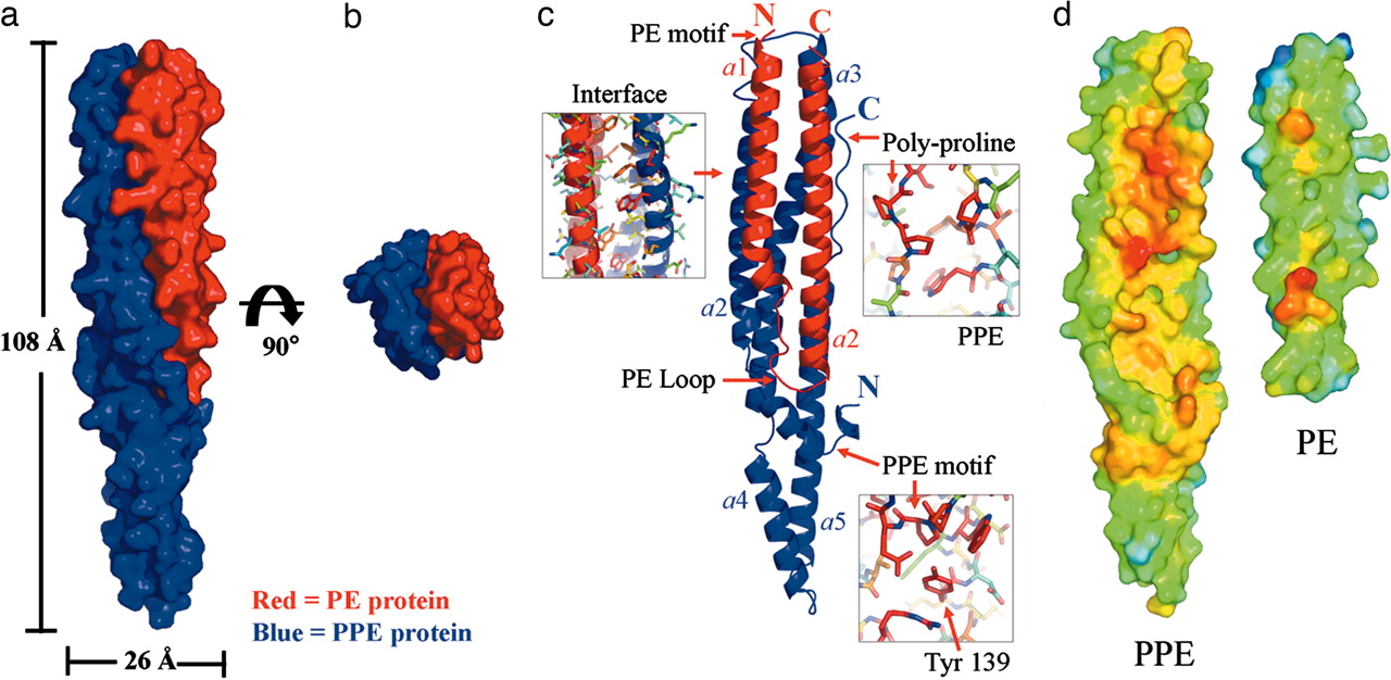
| 14:40, 8 October 2008 | Strong_PNAS_PE_PPE_2.jpg (file) | Michael Strong | 295 KB | (PE PPE Figure Adapted from Strong et al. Proc Natl Acad Sci U S A. 2006 103: 8060–8065. ) |
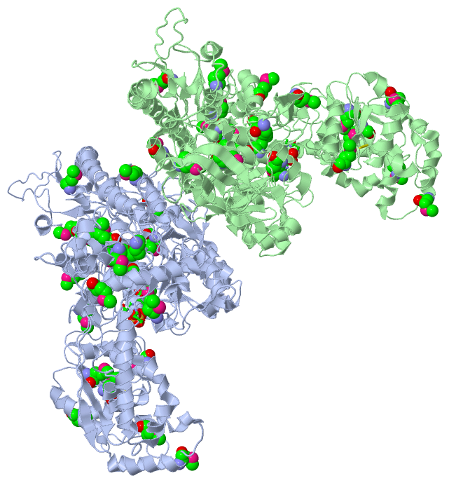
| 14:18, 8 October 2008 | Strong_PNAS_PE_PPE_1.jpg (file) | Michael Strong | 181 KB | (Overview of the PE PPE complex, adapted from Strong et al. Proc Natl Acad Sci U S A. 103:8060-5 (2006).) |
| 15:01, 7 October 2008 | 2OUIal.pdb (file) | Alexander Berchansky | 1.68 MB | |
| 11:15, 7 October 2008 | 2NVBal3.pdb (file) | Alexander Berchansky | 1.65 MB | |
| 10:59, 7 October 2008 | 2NVBal2.pdb (file) | Alexander Berchansky | 1.66 MB | |
| 10:51, 7 October 2008 | 2NVBal1.pdb (file) | Alexander Berchansky | 1.74 MB | |
| 09:46, 7 October 2008 | 2NVBal.pdb (file) | Alexander Berchansky | 1.74 MB | |
| 23:52, 6 October 2008 | Newsplat.gif (file) | Eric Martz | 386 B | |
| 20:32, 5 October 2008 | Lacrep_anim_small.gif (file) | Eric Martz | 98 KB | (Multi-gif animation of morph of the lac repressor bound to DNA, changing from non-specific to specific recognition. [http://www.umass.edu/microbio/chime/pe_beta/pe/atlas/morphs/lacrep/index.htm Details]) |
| 03:09, 5 October 2008 | 1osl_19_1l1m_9_morph.pdb (file) | Eric Martz | 451 KB | (Linear interpolation morph of 1osl model 19 to 1L1M model 9. Lac repressor binding to DNA, going from nonspecific binding to specific binding.) |
| 17:41, 3 October 2008 | 2chg9-63_aligned_with_dnac_model.pdb (file) | Eric Martz | 126 KB | (See User:Eric Martz/Sandbox 4) |
| 20:08, 2 October 2008 | Dnac_from_2ggz_a.pdb (file) | Eric Martz | 98 KB | (DnaC gene of E coli, sequence homology modeled by Swiss Model using template 2ggz:A.) |
| 13:06, 2 October 2008 | Sgap1.png (file) | Harry Greenblatt | 366 KB | (View showing PCI-1 bound in the active site of SGPB showing surface of interaction on enzyme) |
| 01:43, 29 September 2008 | 1yln.png (file) | OCA | 146 KB | (Source Jena Library http://www.fli-leibniz.de/IMAGE.html) |
| 15:36, 28 September 2008 | 1yud.png (file) | OCA | 458 KB | (Source Jena Library http://www.fli-leibniz.de/IMAGE.html) |
| 14:14, 28 September 2008 | 1tsj.png (file) | OCA | 166 KB | (Source Jena Library http://www.fli-leibniz.de/IMAGE.html) |
| 13:47, 28 September 2008 | 2fcj.png (file) | OCA | 313 KB | (Source Jena Library http://www.fli-leibniz.de/IMAGE.html) |
| 00:42, 28 September 2008 | Active_site_still.png (file) | Tom Gluick | 37 KB | (The image shows the active site residues of Chain A of human glutamine synthetase.) |
| 06:08, 24 September 2008 | 2pne.png (file) | OCA | 97 KB | (Source Jena Library http://www.fli-leibniz.de/IMAGE.html) |
| 19:33, 23 September 2008 | 2hu4_1.pdb (file) | Eric Martz | 1,020 KB | (One of the two tetramers in the asymmetric unit of 2hu4. Obtained as one of the biological units from pdb.org.) |
| 14:38, 23 September 2008 | 2B83a.pdb (file) | Alexander Berchansky | 1.71 MB | (Alignment 1jqb on 2b83) |
| 11:40, 23 September 2008 | Note.gif (file) | Jaime Prilusky | 304 B | |
| 02:44, 23 September 2008 | 2hty2hu4_j.pdb (file) | Eric Martz | 446 KB | (Linear interpolation morph from chain A of 2hty to chain A of 2huj with 6 intermediate interpolated models. The ligand G39 (Tamiflu) has been included in all 8 models, even though it is meaningful only in model 8 (chain A of 2huj). This was done so its po) |
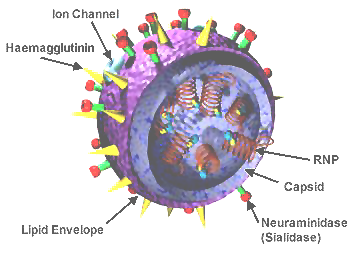
| 22:14, 21 September 2008 | 3D_Influenza_virus.png (file) | Eric Martz | 79 KB | (Obtained from wikipedia, http://en.wikipedia.org/wiki/Image:3D_Influenza_virus.png) |
| 18:43, 14 September 2008 | TRACE_SF_LABEL_2QC8A.jpg (file) | Tom Gluick | 24 KB | (This file shows the Chain A with ligands from human glutamine synthetase, PDB 2qc8) |
| 15:39, 14 September 2008 | 1STF_2.pdb (file) | Nir London | 442 KB | |
| 15:38, 14 September 2008 | 1STF_1.pdb (file) | Nir London | 203 KB | |
| 15:31, 14 September 2008 | 1STF_true.pdb (file) | Nir London | 203 KB | (This is a CORRECT docking model of the complex from 1stf) |
| 15:30, 14 September 2008 | 1STF_false.pdb (file) | Nir London | 442 KB | (This is a WRONG docking model of the complex from 1stf) |
| 15:11, 14 September 2008 | 1STF.pdb (file) | Nir London | 442 KB | (a WRONG model of the complex in 1STF pdb, part of the FunHunt training set) |
| 07:26, 9 September 2008 | 1XXM.pdb (file) | Alexander Berchansky | 299 KB |
First page |
Previous page |
Next page |
Last page |
