File list
From Proteopedia
| Name | User | Size | Description | |
|---|---|---|---|---|
| 07:39, 20 February 2010 | Moran_asite_par_morph.pdb (file) | Eric Martz | 315 KB | |
| 23:53, 19 February 2010 | GFP_Chromophore.png (file) | Laura Carbone | 71 KB | (Self-catalyzed cyclization and oxidation of the GFP chromophore) |
| 13:20, 18 February 2010 | 7f6d0227c1086977a55e1a6ae41c5afa_pdb1.pdb (file) | OCA | 411 KB | (converted) |
| 13:19, 18 February 2010 | Cfb0c29e1d660bd6780db20d1ca56aab_pdb1.pdb (file) | OCA | 45 KB | (converted) |
| 13:18, 18 February 2010 | A6f0c902ee4c01b663ec2c214b093436_pdb1.pdb (file) | OCA | 411 KB | (converted) |
| 13:05, 18 February 2010 | Db5b95958bad4d4cb0f79aaa22bc33bc_pdb1.pdb (file) | OCA | 411 KB | (converted) |
| 13:03, 18 February 2010 | 37b839a0dbe021cb0cbb10f79d388e50_pdb1.pdb (file) | OCA | 411 KB | (converted) |
| 11:04, 18 February 2010 | Kyc.pdb (file) | Alexander Berchansky | 1.66 MB | (Alignment of 3kyd & 1y8r on 3kyc (chains A)) |
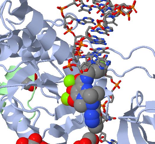
| 00:43, 18 February 2010 | Strand_Inhibition.jpg (file) | Jordan Heard | 60 KB | (JMOL image of MK-0518 (Raltegravir) blocking the 3' end of the viral DNA from binding the active site. Notice the interaction between the metal chelating oxygen atoms on MK-0518 and the magnesium cations present in the active site.) |
| 00:31, 18 February 2010 | Rnl2_Electorstatic_potential.jpg (file) | William Eisbrenner | 1.34 MB | |
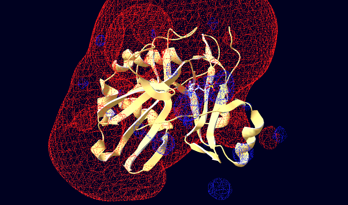
| 21:51, 17 February 2010 | Pockets_global.jvxl (file) | Karl Oberholser | 42 KB | (surface made by Jmol command isosurface minset 1000 ignore (A8S or HOH) POCKET sasurface 0.5 with all protein selected.) |
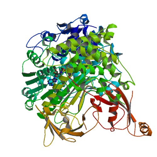
| 08:51, 17 February 2010 | 3kyc.jpg (file) | OCA | 66 KB | (Source PDB http://www.rscb.org/pdb) |
| 23:43, 16 February 2010 | 3hg3to4.pdb.gz (file) | Wayne Decatur | 957 KB | (morph of 3hg3 to 3hg4) |
| 22:46, 16 February 2010 | Better3hg2to3.pdb.gz (file) | Wayne Decatur | 963 KB | (3hg2 to 3hg3 morph) |
| 22:44, 16 February 2010 | 3hg2to3.pdb.gz (file) | Wayne Decatur | 963 KB | (added in the molecule b substrate) |
| 19:10, 16 February 2010 | 3fpntest.pdb (file) | Wayne Decatur | 260 KB | (test of 3fpn multimodel with correct model markers) |
| 18:39, 16 February 2010 | Pocket_included_A8S_HOH.jvxl (file) | Karl Oberholser | 7 KB | (Surface of binding pocket of abscisic acid in chain a only, 3k3k.pdb. The jvxl file was generated by selecting the residues making up the AC1 site and not ignoring A8S & HOH.) |
| 15:20, 16 February 2010 | Two_binding_pockets.jvxl (file) | Karl Oberholser | 11 KB | (Surfaces of binding pockets of abscisic acid in its receptor, 3k3k.pdb. The surfaces (jvxl file) were generated by selecting the residues making up the AC1 site and using the Jmol command: isosurface minset 1000 ignore (A8S or HOH) POCKET cavity 1.0 sasu) |
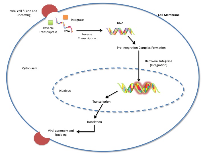
| 04:15, 14 February 2010 | GAViralLifeCycle.jpg (file) | Garrett Asanuma | 59 KB | |
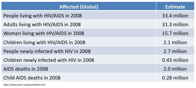
| 03:27, 14 February 2010 | HIV_Stats.jpg (file) | Garrett Asanuma | 63 KB | |
| 09:04, 11 February 2010 | ADH3a.pdb (file) | Alexander Berchansky | 628 KB | |
| 09:03, 11 February 2010 | ADH2a.pdb (file) | Alexander Berchansky | 820 KB | |
| 08:36, 11 February 2010 | ADH3.pdb (file) | Alexander Berchansky | 628 KB | (Alignment of 1ped, 1y9a, and 3fpc) |
| 08:11, 11 February 2010 | ADH2.pdb (file) | Alexander Berchansky | 820 KB | (Alignment 3ftn (A,B) on 3fsr (A,B)) |
| 07:40, 11 February 2010 | Fsrab.pdb (file) | Alexander Berchansky | 820 KB | (ALignment of 3ftn on 3fsr (chains A,B)) |
| 14:53, 10 February 2010 | Fsrad.pdb (file) | Alexander Berchansky | 832 KB | (Alignment of 3ftn (chain A, D) on 3fsr (chain A, D) ) |
| 11:48, 10 February 2010 | ADH1.pdb (file) | Alexander Berchansky | 1.03 MB | (Alignment of chain A of 1ped on chain A of 3fsr) |
| 10:11, 10 February 2010 | 115808f4d66c3b21802e9ae07e45112c_pdb1.pdb (file) | OCA | 965 KB | (converted) |
| 16:11, 9 February 2010 | 456150b98a2ddf2cce223fd369d1144d_pdb1.pdb (file) | OCA | 11 KB | (converted) |
| 16:11, 9 February 2010 | 456150b98a2ddf2cce223fd369d1144d_jmol.spt (file) | OCA | 3 KB | (converted) |
| 16:10, 9 February 2010 | B5b3e16b5c25745d812fccf9301fd860_pdb1.pdb (file) | OCA | 11 KB | (converted) |
| 16:10, 9 February 2010 | B5b3e16b5c25745d812fccf9301fd860_jmol.spt (file) | OCA | 3 KB | (converted) |
| 15:26, 9 February 2010 | D4154567ae36d2470abcf0446195080e_pdb1.pdb (file) | OCA | 962 KB | (converted) |
| 15:26, 9 February 2010 | D4154567ae36d2470abcf0446195080e_jmol.spt (file) | OCA | 61 KB | (converted) |
| 09:33, 8 February 2010 | Kk.pdb (file) | Student | 36 KB | |
| 09:17, 8 February 2010 | Finaltet.pdb (file) | Student | 315 KB | (Gse1 molecule) |
| 22:18, 7 February 2010 | GFP_mice.png (file) | Laura Carbone | 115 KB | |
| 13:56, 6 February 2010 | Jmol_edm_tyr_1.0s.png (file) | Eric Martz | 28 KB | (Electron density map from 3hyd.) |
| 13:07, 31 January 2010 | Quiz_feedback02.png (file) | Eric Martz | 18 KB | (snapshot for Help:Quiz) |
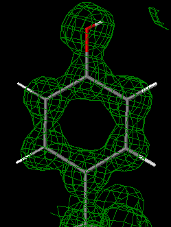
| 12:59, 28 January 2010 | 3hf1.png (file) | OCA | 407 KB | (Source Jena Library http://www.fli-leibniz.de/IMAGE.html) |
| 17:57, 27 January 2010 | 3ftn.jpg (file) | OCA | 67 KB | (Source PDB http://www.rscb.org/pdb) |
| 04:21, 26 January 2010 | 3jyxwvALPHAC.pdb.gz (file) | Wayne Decatur | 256 KB | (This structure is a composite of 3 pdb files optimized for use online: 3jyx is a 3 chains structure of sequences from Thermomyces lanuginosus. 3jyw is a 30 chains structure of sequences from Thermomyces lanuginosus. 3jyv is a 19 chains structure of sequ) |
| 17:32, 22 January 2010 | 2H24.pdb (file) | Shelly Huebert | 129 KB | |
| 13:39, 20 January 2010 | 3fsr.jpg (file) | OCA | 67 KB | (Source PDB http://www.rscb.org/pdb) |
| 13:38, 20 January 2010 | 3fpl.jpg (file) | OCA | 93 KB | (Source PDB http://www.rscb.org/pdb) |
| 13:37, 20 January 2010 | 3fpc.jpg (file) | OCA | 68 KB | (Source PDB http://www.rscb.org/pdb) |
| 21:52, 18 January 2010 | 1DG5.pdb (file) | Joseph Kinscher | 151 KB | (DHFR) |
| 15:45, 15 January 2010 | FIH_activesite.PDB (file) | John Hangasky | 8 KB | |
| 15:08, 13 January 2010 | UMP.pdb (file) | Karl Oberholser | 2 KB | |
| 14:53, 13 January 2010 | CMP.pdb (file) | Karl Oberholser | 2 KB |
First page |
Previous page |
Next page |
Last page |
