This old version of Proteopedia is provided for student assignments while the new version is undergoing repairs. Content and edits done in this old version of Proteopedia after March 1, 2026 will eventually be lost when it is retired in about June of 2026.
Apply for new accounts at the new Proteopedia. Your logins will work in both the old and new versions.
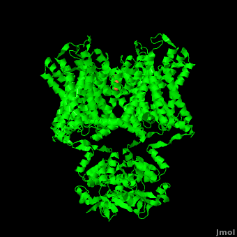
Potassium Channel
From Proteopedia
| |||||||||||

Page Development
This article was developed based on lectures given in Chemistry 543 by Prof. Clarence E. Schutt at Princeton University.
Additional Structures of Potassium Channels
Update June 2011
Potassium channels (KCh) are subdivided into voltage-gated KCh and calcium-dependent KCh. The latter are subdivided into high- (BK, LKCa), intermediate- and small-conductance KCh (human SK1, rat SK2, SKCa). The T1 domain is a highly conserved N-terminal domain which is responsible for driving the tetramerization of the KCh α subunit. The inward rectifier KCh (IRK) passes current more easily in the inward direction. MthK is a calcium-dependent KCh from Methanobacterium thermoautrophicum.
1wlj, 2wlh, 2wli, 2wlj, 2wlm, 2wln, 2wlo - MmKCh - Magnetospirillum magnetoacticum
2k44 – KCh voltage-sensor paddle domain – NMR
3e86, 3e83 – BcKCh transmembrane domain – Bacillus cereus
3e8g, 3e89, 3e8b, 3e8h – BcKCh transmembrane domain +ions
2q67, 2q68, 2q69, 2q6a, 3ouf – BcKCh (mutant)
2ahy, 2ahz – BcKCh+ions
1bl8, 1f6g - SlKCh (mutant) - Streptomyces lividans
2qto - SlKCh
1j95 - SlKCh+K+tetrabutylammonium
1jvm - SlKCh (mutant)+Rb+tetrabutylammonium
1jq1, 1jq2 - SlKCh inner transmembrane segment - NMR
3ifx - SlKCh pore domain
2pnv – rKCh leucine zipper domain SKCa – rat
3lut - rKCh Kv1.2 (mutant)
1dsx - rKCh N-terminal (mutant)
1qdv, 1qdw - rKCh Kv1.2 N-terminal
1kn7 - rKCh Kv1.4 N-terminal (mutant) - NMR
1nn7 - rKCh Kv4.2 N-terminal T1 domain
3eau - rKCh beta2 +cortisone
3eb3, 3eb4 - rKCh beta2 (mutant) + cortisone
1a68, 1eod, 1eoe, 1eof, 3kvt - AcKCh Kv1.1 T1 domain (mutant) - Aplysia californica
1t1d - AcKCh Kv1.1 T1 domain
1b4g - hKCh inactivation domain - human - NMR
1byw - hKCh ERG N-terminal (mutant)
1ujl - hKCh ERG1 extracellular linker - NMR
2l1m, 2l4r - hKCh HERG - NMR
1s1g - hKCh Kv4.3 T1 domain
2ovc - hKCh Kv7.4 T1 domain
3bj4 - hKCh Kv7.1 C-termianl
3hfc, 3hfe - hKCh Kv7.1 (mutant)
1zxs - hKCh beta2
3co2, 1vp6 - MlotiK1 cyclic nucleotide binding domain - Mesorhizobium loti
1ho2, 1ho7 - KCh L45 segment - Drosophila melanogaster - NMR
2kyh - AeKCh voltage ensing domain - Aeropyrum pernix - NMR
2wll - KCh KIRBAC1.1 - Burkholderia pseudomalie
Potassium channel complex with protein
2nz0 – hKCh Kv4.3 N-terminal+KV channel interacting protein 1
2i2r - rKCh Kv4.3 N-terminal+KV channel interacting protein 1
1s6c - rKCh Kv4.2 N-terminal+KV channel interacting protein 1
2a79, 1qrq - rKCh Kv1.2 + beta2
2r9r - rKCh
1qx7 – rKCh SKCa+ calmodulin
1g4y – rKCh rSK2 calmodulin binding domain SKCa + calmodulin
1exb - rKCh Kv1.1 T1 domain + KV beta2 protein
2p7t - rKCh + FAV
3lnm - rKCh Kv2.1/KCh Kv1.2 (mutant)
3eff - mKCh + FAB - mouse
2w0f - mKCh + FAB + tetraoctylammonium
2hg5, 2h8p, 2hfe - KCh + mFAB
3f7v, 3f7y, 3fb5, 3fb7, 3fb8, 3gb7, 3hpl, 3iga, 3or6, 3or7, 1r3i, 1r3j, 1r3k,1r3l, 1s5h, 2atk, 2bob, 2boc, 2dwd, 2dwe, 2hvj, 2hvk, 2ih1, 2ih3, 2itc, 2itd, 2jk5, 2nlj - mKCh (mutant) + FAB
3f5w, 1zwi, [2hjf]] - mKCh (mutant) + antibody heavy+light chains
1k4c, 1k4d - SlKCh (mutant) + antibody heavy+light chains
2a0l - AeKCh + FV
1orq - AeKCh (mutant) + FAB
2a9h - SlKCh (mutant) + charybdotoxin
Inward rectifier KCh
1u4f,3agw - mIRK 2 cytoplasmic domain
2gix - mIRK 2 cytoplasmic domain (mutant)
2e4f - mIRK 2 fragment
1n9p, 1u4e - mIRK 1 cytoplasmic domain
3k6n - mIRK 1 cytoplasmic domain (mutant)
1xl4, 1xl6, 2wlk, 2x6a, 2x6b, 2x6c - MmIRK KIRBAC3.1
1p7b - BpIRK C-terminal
MthK
1kxd - MthK RCK domain + Cd - Methanobacterium thermoautrophicum
2ogu, 2fy8, 2aej, 2aem, 1lnq - MthK RCK domain
3ldc, 3ldd, 3lde, 3ous – MthK residues 28-99 (mutant)
2aef, 3kxd - MthK RCK domain + Ca
BK channel
3mt5 - hBK cytoplasmic domain
1jo6 - BK beta 2 N-terminal KCNMB2 encoded LKCa - NMR
Potassium/Sodium channel
3k03, 3k04, 3k06, 3k08, 3k0d, 3k0g – BcNaK
Calcium-activated KCh
3naf – hKCh α1 subunit
Additional Resources
For Additional Information, See: Membrane Channels & Pumps
References
- ↑ 1.0 1.1 Zhou Y, Morais-Cabral JH, Kaufman A, MacKinnon R. Chemistry of ion coordination and hydration revealed by a K+ channel-Fab complex at 2.0 A resolution. Nature. 2001 Nov 1;414(6859):43-8. PMID:11689936 doi:http://dx.doi.org/10.1038/35102009
- ↑ 2.0 2.1 2.2 2.3 Doyle DA, Morais Cabral J, Pfuetzner RA, Kuo A, Gulbis JM, Cohen SL, Chait BT, MacKinnon R. The structure of the potassium channel: molecular basis of K+ conduction and selectivity. Science. 1998 Apr 3;280(5360):69-77. PMID:9525859
- ↑ Jiang Y, Lee A, Chen J, Ruta V, Cadene M, Chait BT, MacKinnon R. X-ray structure of a voltage-dependent K+ channel. Nature. 2003 May 1;423(6935):33-41. PMID:12721618 doi:http://dx.doi.org/10.1038/nature01580
- ↑ Waters MF, Minassian NA, Stevanin G, Figueroa KP, Bannister JP, Nolte D, Mock AF, Evidente VG, Fee DB, Muller U, Durr A, Brice A, Papazian DM, Pulst SM. Mutations in voltage-gated potassium channel KCNC3 cause degenerative and developmental central nervous system phenotypes. Nat Genet. 2006 Apr;38(4):447-51. Epub 2006 Feb 26. PMID:16501573 doi:ng1758
- ↑ 5.0 5.1 5.2 5.3 Long SB, Tao X, Campbell EB, MacKinnon R. Atomic structure of a voltage-dependent K+ channel in a lipid membrane-like environment. Nature. 2007 Nov 15;450(7168):376-82. PMID:18004376 doi:http://dx.doi.org/10.1038/nature06265
Proteopedia Page Contributors and Editors (what is this?)
Michal Harel, David Canner, Joel L. Sussman, Alexander Berchansky
