Zinc Fingers
From Proteopedia

|

Contents |
Overview
Over the course of time, natural selection has produced multiple solutions for the optimization of gene transcription, and few other transcription factors highlight this fact better than the Zing Finger Proteins (ZFPs). While there are other zinc-containing subregions of other proteins, the distinguishing feature of a zinc finger is spontaneous folding via coordination of a zinc ion, despite is its small size. They are highly specific to their sequence and yet are able to target sequences common to multiple loci for regulation of a common function that may be influenced by many genes. They can also serve as carrier to other domains which may bind covalently to the DNA, effecting permanent shutdown of a gene’s expression. A true testament to their importance in the human genome, a family of over 700 human proteins contain a Zinc Finger domain, a number exceeded only the immunoglobins, and then only slightly.
Basic Structural Profile of a Zinc Finger
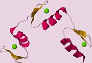
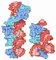
The simplest group of zinc fingers, referred as C2H2 zinc fingers, consists of two antiparallel β-pleated sheets and a right-handed α-helix. The name C2H2, or Cis2His2, gives a nod to the two cysteine and two histidine residues involved in coordination of the zinc ion. The turn between the two β-pleated sheets forms a hydrophobic pocket near where the zinc ion is bound. Typically, the hydrophobic pocket is formed as a result of interactions between phenylalanines and leucines, in the close proximity. While both the coordination of the zinc ligand and the presence of the hydrophobic pocket stabilize the small zinc finger domain, the zinc ligand is responsible for a majority of the stability imparted to the motif. Because cells contain a highly reducing environment, sulfide bridges are unable to stabilize small protein domains. With a single oxidation state and the ability to accommodate both nitrogen and sulfur, Zinc is an ideal stabilizer. Due to the structural stability imparted to the motif by the zinc ion, zinc fingers are considerably smaller than most other proteins, usually ranging between 25 and 30 amino acids in length. As a corollary of their small size, they are extremely agile and mobile, and so are the genes that encode them. Thus, they are easily able to bind to DNA, among other substrates. By slipping into the major groove of DNA, they are able to use their amino acids to check for proper base identity. This allows the zinc fingers to bind to sequences normally unavailable to other, larger DNA-binding structural motifs. As a family, the structure of the zinc fingers is as polymorphous as it is unique. Some are coordinated primarily or exclusively by cysteine, and many form much more complex structures than the β-hairpin of Cis2His2 types.
Biological Role and Regulation
Given the prevalence of the zinc fingers in nature, it is no surprise to learn that they are key players in many biological processes. Gli-1, a zinc-finger transcription factor involved in embryonic development, is also implicated in carcinogenesis when it is over-expressed in dividing cells. Likewise, Gli-3 is another zinc-finger transcription factor whose shortened repressor form has roles in modulating regulation of genes controlling apoptosis, and consequently has a dramatic effect on development of the limb bud and the digit formation that follows. In part, the regulation of the zinc fingers is kept in check by other repressors or activators, as well as various types of chromatin remodeling (e.g. acetylation, methylation) which definitively blocks transcription of their targets.
Clinical Applications
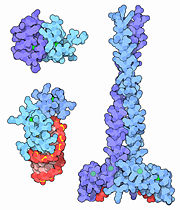
The advantages of using ZFPs in a clinical setting are numerous, the least of which is not their ability to bind DNA. As a result, the ZFP must bind two only two copies of its target, as opposed to therapeutics directed at mRNA, for example, which have to are in direct quantitative competition with their target. Because of this unique advantage as well as their modularity and ability to work in clusters, the ZFPs are currently the subject of extensive research for use in genetic therapy. Their versatility and modularity among targets can be explained as a function of their α-helical sidechains, which they use to interact electrostatically with multiple sequences. Because they can withstand multiple mutations without losing their functional structure, they make great candidates for specific gene-targeting applications. In theory, the zinc finger as a therapeutic agent could unilaterally control gene expression, given that a transcription factor had been synthesized which possessed the proper sequence identity for binding to the gene or the regulated protein.
3D Structure of Zinc Finger Domains
1hvo
1x68
1x6a
1x6f
2bai
2ctu
2ds6
2e72
2epc
2epp
2epq
2epr
2eps
2ept
2epu
2epv
2epw
2epx
2epy
2epz
2eq0
2eq1
2eq2
2eq3
2eq4
2rpp
2ysv
Additional Information
- Zinc Finger in Wikipedia
- Zinc Finger Consortium
- Molecule of the Month on Zinc Fingers by David Goodsell
- Molecule of the Month at Teaching Scenes, Tutorials, and Educators' Pages
- For Additional Information, See: Transcription
Content Donators
Many images on this page are the work of David S. Goodsell, who has given permission for their inclusion in Proteopedia:
- Content adapted with permission from David S. Goodsell's Molecule of the Month on Zinc Fingers
Proteopedia Page Contributors and Editors (what is this?)
Ala Jelani, Ann Taylor, Eran Hodis, Michal Harel, Tyler Combs, Joel L. Sussman, David Canner, Eric Martz

