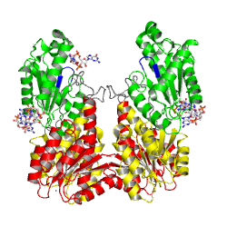
Response regulator PLED in complex with C-di-GMP
From Proteopedia
Contents |
RESPONSE REGULATOR PLED IN COMPLEX WITH C-DI-GMP
Phosphorylation site:
Substrate binding site:
with c-di-GMP bound
Note that the ligand is cross-linking to another substrate binding site
(belonging to an adjacent dimer, not shown).
Recent discoveries suggest that a novel second messenger, bis-(3'-->5')-cyclic di-GMP (c-diGMP), is extensively used by bacteria to control multicellular behavior. Condensation of two GTP to the dinucleotide is catalyzed by the widely distributed diguanylate cyclase (DGC or GGDEF) domain that occurs in various combinations with sensory and/or regulatory modules. The crystal structure of the unorthodox response regulator PleD from Caulobacter crescentus, which consists of two CheY-like receiver domains and a DGC domain, has been solved in complex with the product c-diGMP. PleD forms a dimer with the CheY-like domains (the stem) mediating weak monomer-monomer interactions. The fold of the DGC domain is similar to adenylate cyclase, but the nucleotide-binding mode is substantially different. The guanine base is H-bonded to Asn-335 and Asp-344, whereas the ribosyl and alpha-phosphate moieties extend over the beta2-beta3-hairpin that carries the GGEEF signature motif. In the crystal, c-diGMP molecules are crosslinking active sites of adjacent dimers. It is inferred that, in solution, the two DGC domains of a dimer align in a two-fold symmetric way to catalyze c-diGMP synthesis. Two mutually intercalated c-diGMP molecules are found tightly bound at the stem-DGC interface. This allosteric site explains the observed noncompetitive product inhibition. We propose that product inhibition is due to domain immobilization and sets an upper limit for the concentration of this second messenger in the cell.
Structural basis of activity and allosteric control of diguanylate cyclase., Chan C, Paul R, Samoray D, Amiot NC, Giese B, Jenal U, Schirmer T, Proc Natl Acad Sci U S A. 2004 Dec 7;101(49):17084-9. Epub 2004 Nov 29. PMID:15569936
From MEDLINE®/PubMed®, a database of the U.S. National Library of Medicine.
About this Structure
1W25 is a Single protein structure of sequence from Caulobacter vibrioides. Full crystallographic information is available from OCA.
Also see C-di-GMP_signaling#PleD.
Primary Reference
Structural basis of activity and allosteric control of diguanylate cyclase., Chan C, Paul R, Samoray D, Amiot NC, Giese B, Jenal U, Schirmer T, Proc Natl Acad Sci U S A. 2004 Dec 7;101(49):17084-9. Epub 2004 Nov 29. PMID:15569936
Further References
Activated PleD structure 2v0n:
- Wassmann P, Chan C, Paul R, Beck A, Heerklotz H, Jenal U, Schirmer T. Structure of BeF3- -modified response regulator PleD: implications for diguanylate cyclase activation, catalysis, and feedback inhibition. Structure. 2007 Aug;15(8):915-27. PMID:17697997 doi:http://dx.doi.org/10.1016/j.str.2007.06.016
- Paul R, Weiser S, Amiot NC, Chan C, Schirmer T, Giese B, Jenal U. Cell cycle-dependent dynamic localization of a bacterial response regulator with a novel di-guanylate cyclase output domain. Genes Dev. 2004 Mar 15;18(6):715-27. PMID:15075296 doi:http://dx.doi.org/10.1101/gad.289504
- Paul R, Abel S, Wassmann P, Beck A, Heerklotz H, Jenal U. Activation of the diguanylate cyclase PleD by phosphorylation-mediated dimerization. J Biol Chem. 2007 Oct 5;282(40):29170-7. Epub 2007 Jul 19. PMID:17640875 doi:http://dx.doi.org/10.1074/jbc.M704702200
Created by Tilman Schirmer.
Categories: Caulobacter vibrioides | Single protein | Chan, C. | Jenal, U. | Schirmer, T. | Allosteric product inhibition | Cyclic dinucleotide | Cyclic-digmp | Cyclic-di-GMP | C-di-GMP | Diguanylate cyclase | Ggdef domain | Phosphorylation | Response regulator | Signaling protein | Two-component system

