This old version of Proteopedia is provided for student assignments while the new version is undergoing repairs. Content and edits done in this old version of Proteopedia after March 1, 2026 will eventually be lost when it is retired in about June of 2026.
Apply for new accounts at the new Proteopedia. Your logins will work in both the old and new versions.
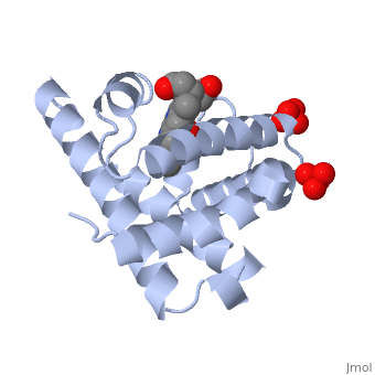
1a6m
From Proteopedia

| |||||||||
| 1a6m, resolution 1.00Å () | |||||||||
|---|---|---|---|---|---|---|---|---|---|
| Ligands: | , , | ||||||||
| |||||||||
| |||||||||
| |||||||||
| Resources: | FirstGlance, OCA, PDBsum, RCSB | ||||||||
| Coordinates: | save as pdb, mmCIF, xml | ||||||||

Contents |
OXY-MYOGLOBIN, ATOMIC RESOLUTION
Template:ABSTRACT PUBMED 10512835
About this Structure
1a6m is a 1 chain structure of Myoglobin with sequence from Physeter catodon. Full crystallographic information is available from OCA.
See Also
Reference
- Vojtechovsky J, Chu K, Berendzen J, Sweet RM, Schlichting I. Crystal structures of myoglobin-ligand complexes at near-atomic resolution. Biophys J. 1999 Oct;77(4):2153-74. PMID:10512835 doi:http://dx.doi.org/10.1016/S0006-3495(99)77056-6
- Bhattacharyya R, Samanta U, Chakrabarti P. Aromatic-aromatic interactions in and around alpha-helices. Protein Eng. 2002 Feb;15(2):91-100. PMID:11917145
- Chen X, Weber I, Harrison RW. Hydration water and bulk water in proteins have distinct properties in radial distributions calculated from 105 atomic resolution crystal structures. J Phys Chem B. 2008 Sep 25;112(38):12073-80. Epub 2008 Aug 28. PMID:18754631 doi:10.1021/jp802795a
