2rh1
From Proteopedia
| |||||||||
| 2rh1, resolution 2.40Å () | |||||||||
|---|---|---|---|---|---|---|---|---|---|
| Ligands: | , , , , , , , | ||||||||
| Gene: | ADRB2, ADRB2R, B2AR / E (Enterobacteria phage T4) | ||||||||
| |||||||||
| |||||||||
| Resources: | FirstGlance, OCA, RCSB, PDBsum | ||||||||
| Coordinates: | save as pdb, mmCIF, xml | ||||||||
Contents |
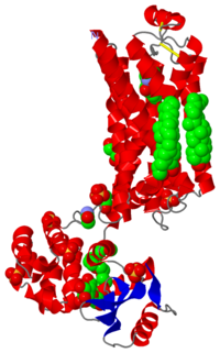
High resolution crystal structure of human B2-adrenergic G protein-coupled receptor.
Heterotrimeric guanine nucleotide-binding protein (G protein)-coupled receptors constitute the largest family of eukaryotic signal transduction proteins that communicate across the membrane. We report the crystal structure of a human beta2-adrenergic receptor-T4 lysozyme fusion protein bound to the partial inverse agonist carazolol at 2.4 angstrom resolution. The structure provides a high-resolution view of a human G protein-coupled receptor bound to a diffusible ligand. Ligand-binding site accessibility is enabled by the second extracellular loop, which is held out of the binding cavity by a pair of closely spaced disulfide bridges and a short helical segment within the loop. Cholesterol, a necessary component for crystallization, mediates an intriguing parallel association of receptor molecules in the crystal lattice. Although the location of carazolol in the beta2-adrenergic receptor is very similar to that of retinal in rhodopsin, structural differences in the ligand-binding site and other regions highlight the challenges in using rhodopsin as a template model for this large receptor family.
High-resolution crystal structure of an engineered human beta2-adrenergic G protein-coupled receptor., Cherezov V, Rosenbaum DM, Hanson MA, Rasmussen SG, Thian FS, Kobilka TS, Choi HJ, Kuhn P, Weis WI, Kobilka BK, Stevens RC, Science. 2007 Nov 23;318(5854):1258-65. Epub 2007 Oct 25. PMID:17962520
From MEDLINE®/PubMed®, a database of the U.S. National Library of Medicine.
About this Structure
2rh1 is a 1 chain structure with sequence from Enterobacteria phage t4. The April 2008 RCSB PDB Molecule of the Month feature on Adrenergic Receptors by David S. Goodsell is 10.2210/rcsb_pdb/mom_2008_4. Full crystallographic information is available from OCA.
See Also
- Adrenergic receptor
- Beta-2 Adrenergic Receptor
- Nobel Prizes for 3D Molecular Structure
- Suggestions for new articles
- User:Wayne Decatur/NASCE2011
- User:Wayne Decatur/UNH BCHEM833 Structural Proteomics Introductory Lecture Fall 2012
Reference
- Cherezov V, Rosenbaum DM, Hanson MA, Rasmussen SG, Thian FS, Kobilka TS, Choi HJ, Kuhn P, Weis WI, Kobilka BK, Stevens RC. High-resolution crystal structure of an engineered human beta2-adrenergic G protein-coupled receptor. Science. 2007 Nov 23;318(5854):1258-65. Epub 2007 Oct 25. PMID:17962520
Categories: Adrenergic Receptors | Enterobacteria phage t4 | RCSB PDB Molecule of the Month | ATCG3D, Accelerated Technologies Center for Gene to 3D Structure. | Cherezov, V. | Choi, H J. | GPCR, GPCR Network. | Hanson, M A. | Kobilka, B K. | Kobilka, T S. | Kuhn, P. | Rasmussen, S G.F. | Rosenbaum, D M. | Stevens, R C. | Thian, F S. | Weis, W I. | 7tm | Accelerated technologies center for gene to 3d structure | Adrenergic | Atcg3d | Cholesterol | Fusion | Gpcr | Lipidic | Lipidic cubic phase | Membrane protein | Membrane protein - hydrolase complex | Mesophase | Protein structure initiative | Psi-2 | Structural genomic


