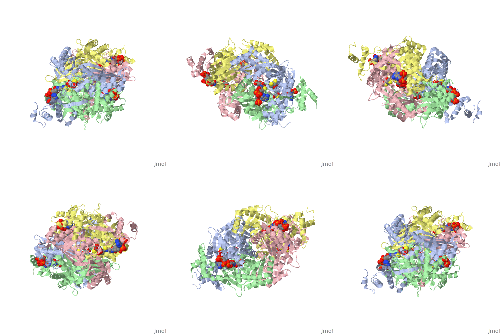
Ligand Binding Domain
The Ligand binding domain for each piece of the dimer has a very similar structure of an .
RARα-RXRα Heterodimer
When these two proteins form a heterodimer, the overall structure of the larger dimer is comparable to that of an RXRα homodimer, likely due to the many similarities these two molecules share. RARα and RXRα rely on residues from the H7, H8, H9, H10, L8-9, and L9-10 domains of both molecules to form the heterodimer interface. The sequence identity between the two molecules on the dimer interface is 0.33, demonstrating that 33% of the interacting residues are homologous between the different proteins. [5]
The residues of RARα that are interacting in the heterodimer are as follows:
Hydrophobic residues: L356, F374, P375, L378, M379, I381 and A389 (yellow);
Negatively charged residues: D338, D349, E353, E357, D383, and E393 (red);
Positively charged residues: K360, R364, H372, K376, K380, and R385 (blue);
Hydrophilic residues: Q315, Q352, T382, and S386 (green).
The residues of RXRα that are interacting in the heterodimer are as follows:
Hydrophobic residues: Y402, P417, F420, A421, L424, L425, L427, P428, A429, and L435 (yellow);
Negatively charged residues: E357, D384, E395, E399, E406, and E439 (red);
Positively charged residues: R353, K361, R398, K410, K422, R426, R431, and K436 (blue);
Hydrophilic residues: S432 (green).
RARα-RXRα Ligand Interactions

